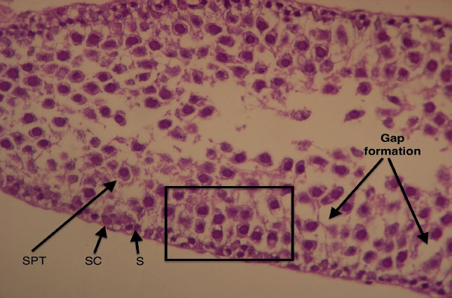
Figure 3. Longitudinal section of a fresh testicular tubule (Group A). The different cells that make up the tubular architecture can be easily made. SC: Sertoli cells; S: spermatogonia cells; SPT: spermatocytes. Formation of intercellular gaps (arrows). See the preservation of tissue architecture in the square (intercellular relation). Magnification 100X.

Figure 4. Longitudinal section of a frozen/thawed testicular tubule (Group B). The different cells that make up the tubular architecture can be easily made. SC: Sertoli cells; S: spermatogonia cells; SPT: spermatocytes. Formation of intercellular gaps (arrows). See the preservation of tissue architecture in the square (intercellular relation). Magnification 100X.
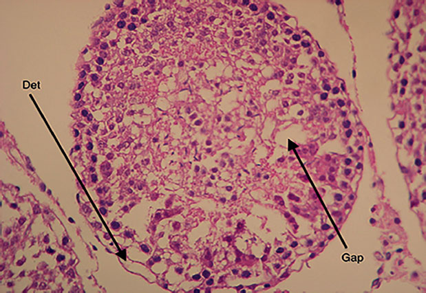
Figure 5. Cross-section of a vitrified/warmed testicular tubule (Group C) showing the formation of intercellular gaps (arrows). Det: detachment from the basement membrane. Magnification 100X.
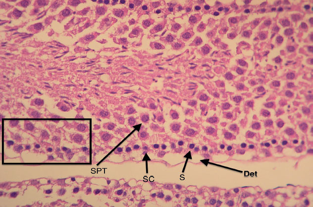
Figure 6. Cross-section of a vitrified/warmed testicular tubule (Group C). The different cells that make up the tubular architecture can be easily made. SC: Sertoli cells; S: spermatogonia cells; SPT; spermatocytes; Det: detachment from the basement membrane. See the preservation of tissue architecture in the square (intercellular relation). Magnification 100X.