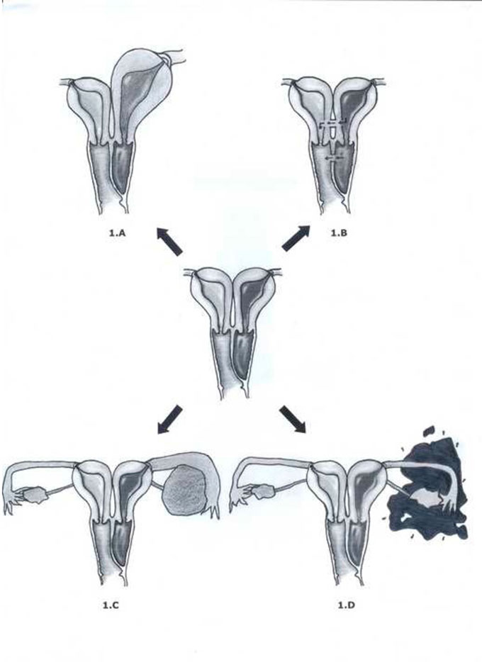
Figure 1 – schematic representation of uterus didelphys with left oblique vaginal septum and hematocolpos adapted from Gidwani & Falcone (1999) with approval.
A,B,C and D - Clinical feature variations according to the different levels of blood retention and complications
Figure 1 A - blood retention with associated tumor formation
Figure 1 B - discrete menstrual discharge through a small puncture
Figure 1 C - tubo-ovarian abscess - TOA (specific case with a 1000 cm3 TOA)
Figure 1 D - blood draining into the peritoneum (increasing the chance of endometriosis)