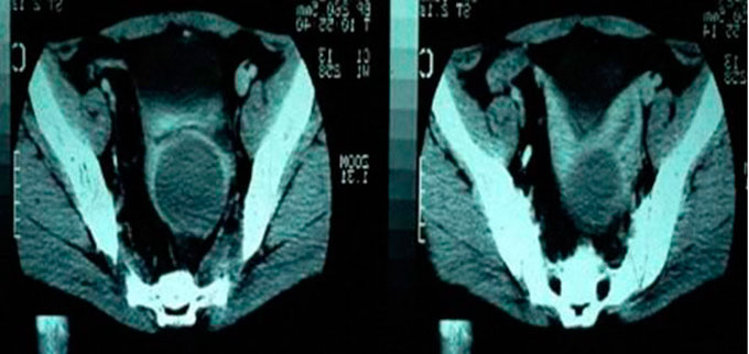
Figure 2 - Magnetic resonance imaging of the pelvis showing uterus didelphys and a large mass of the hematocolpos type in the left hemivagina.
(Case 12 on Table 1)
Figure 2 - Magnetic resonance imaging of the pelvis showing uterus didelphys and a large mass of the hematocolpos type in the left hemivagina.
(Case 12 on Table 1)