JBRA Assist. Reprod. 2015;19 (4):235-240
ORIGINAL ARTICLE
doi: 10.5935/1518-0557.20150046
Endometriosis Affects Oocyte Morphology in Intracytoplasmic Sperm Injection Cycles
¹Fertility – Medical Group – São Paulo – Brasil
²Disciplina de Urologia, Área de Reprodução Humana, Depto de Cirurgia, Universidade Federal de São Paulo
³Instituto Sapientiae – Centro de Estudos e Pesquisa em Reprodução Assistida
CONFLICT OF INTERESTS
No conflict of interest have been declared.
ABSTRACT
Objective: To identify associations between presence of endometriosis and oocyte defects, embryo developmental potential, and cycle outcomes.
Methods: This study looked into the impact of endometriosis on oocyte and embryo quality, and blastocyst formation probability. Endometriosis was also correlated with cycle characteristics. In order to avoid age-related bias, in the first analysis only patients aged 36 years or younger were included, and the cycles were split into endometriosis infertility cycles (n=431; 3172 oocytes) and other cycles (n=2510; 24480 oocytes).
Results: The number of retrieved oocytes (10.6±21.2 vs. 14.6±21.1, P<0.001), oocyte yield (68.1±20.0% vs. 70.6±19.6%, P=0.015), and embryos obtained (6.1±4.43 vs. 7.8±5.12, P<0.001) were lower among patients with endometriosis. Implantation rates (28.1%±38.9% vs. 33.9±42.7, P<0.001) were lower among patients with endometriosis, but fertilization, pregnancy, miscarriage and cycle cancelation rates were not different. There was a significant increase in the incidence of extra-cytoplasmic, but not intra-cytoplasmic, oocyte defects among patients with endometriosis. The quality of embryos (45.3% vs. 47.3%, P=0.037) collected from patients with endometriosis was lower, but blastocyst formation rates were unaltered.
Conclusions: A possible explanation for the lower implantation rates seen in patients with endometriosis is the poorer quality of the oocytes and embryos observed in this group of patients.
Keywords: Endometriosis, ICSI, Oocytes, Embryos, Implantation
INTRODUCTION
It has been suggested that endometriosis is a heterogeneous disease characterized by the presence of endometrial-type cells outside the uterine cavity (Vercellini et al. 2013).
The pathogenesis of endometriosis is still unclear, but retrograde menstruation (Giudice & Kao, 2004), altered immunity (Steele et al., 1984), coelomic metaplasia, metastatic spread (Macer & Taylor, 2012), stem cell and genetic origins have been listed as possible causes (Chan et al., 2004; Du & Taylor, 2007; Zanatta et al., 2010).
Endometriosis patients may be asymptomatic or present a wide variety of symptoms, ranging from pelvic pain to infertility (Burns & Schenken, 1999). Infertility may be present in 30-50% of the individuals with endometriosis (Verkauf, 1987) for unclear reasons. Fecundity is reduced due to mechanical disruptions in cases of advanced pelvic endometriosis with extensive peritoneal adhesions and consequent pelvic anatomy distortions (Holoch & Lessey, 2010).
Substances produced by the implant or adjacent tissues such as prostaglandins, cytokines and growth factors have been listed as possible etiological factors of endometriosis-associated infertility (Olive & Pritts, 2001). Assisted reproductive technologies (ART) constitute a valid alternative for patients with endometriosis-associated infertility (Opoien et al., 2012; Dong et al., 2013).
More than a third of the women offered ART reportedly have endometriosis (Verkauf, 1987); however, studies on the outcome of ART for endometriosis-associated infertility have described conflicting results. Although some studies have reported decreased success rates in minimal/mild endometriosis when compared with non-endometriosis groups (Wardle et al., 1985; Matson & Yovich, 1986; Simon et al., 1994; Arici et al., 1996), other authors have failed to report differences between these groups (Inoue et al., 1992; Olivennes et al., 1995; Tanbo et al., 1995; Meden-Vrtovec et al., 2000). For advanced-stage endometriosis, the results are also contradictory with some reports indicating worse (Meden-Vrtovec et al., 2000; Aboulghar et al., 2003; Kuivasaari et al., 2005; Harb et al., 2013) and others describing comparable outcomes after ART (Al-Fadhli et al., 2006; Gupta et al., 2006; Matalliotakis et al., 2007).
Studies supporting the poor outcome theory suggest that endometriosis-associated infertility patients offered ART present poor ovarian response (Al-Azemi et al., 2000; Matalliotakis et al., 2007; Lin et al., 2010), lower fertilization rates (Gupta et al., 2006; Lin et al., 2010), decreased endometrial receptivity (Giudice & Kao, 2004; Holoch & Lessey, 2010), and poor implantation rates (Harb et al., 2013). It has also been suggested that oocyte and embryo quality (Brizek et al., 1995; Pellicer et al., 1995; Garrido et al., 2000; Ota et al., 2000; Ota et al., 2002; Goud et al., 2014) may be compromised in patients with endometriosis-associated infertility. It has also been reported that the incidence of aneuploidy is significantly higher in patients with endometriosis (Gianaroli et al., 2010).
To our knowledge, the effect of endometriosis-associated infertility on oocyte quality, specifically related to intra- and extra-cytoplasmic defects, has not been investigated yet. Therefore, the goal of this study was to identify associations between the presence of endometriosis and oocyte defects, embryo developmental potential, and cycle outcomes.
MATERIALS AND METHODS
Study Design
This study included oocytes obtained from patients undergoing Intracytoplasmic sperm injection (ICSI) cycles at a private assisted fertilization center between January 2005 and May 2014. The patients were distributed into groups according to the cause of infertility. In order to avoid age bias, only females aged 36 years or younger were included and the cycles were split into endometriosis infertility cycles (n=431; 3172 oocytes) and other cycles (n=2510 cycles; 24480 oocytes).
The endometriosis group included individuals with moderate and severe endometriosis (ASRM III–IV).
The oocytes were evaluated immediately before sperm injection, and the embryos were evaluated 16-18 hours after ICSI and on days two, three and five of development. The influence of the presence of endometriosis on oocyte quality, embryo quality at the cleavage stage (day three), and the rate of blastocyst formation were evaluated. Moreover, the presence of endometriosis was correlated with cycle characteristics and clinical outcomes, such as: (i) number of aspirated follicles; (ii) number of obtained oocytes; (ii) oocyte yield, (iv) mature (MII) oocyte rate, (v) fertilization rate; (vi) clinical pregnancy rate; (vii) miscarriage rate, and (viii) implantation rate.
Patients provided written consent and agreed to share the outcomes of their cycles for research purposes. The local institutional review board approved the study.
Controlled ovarian stimulation
Controlled ovarian stimulation (COS) was achieved by pituitary blockage using a GnRH antagonist (Cetrotide; Serono, Geneva, Switzerland); ovarian stimulation was performed using recombinant FSH (Gonal-F; Serono, Geneva, Switzerland).
Follicular growth was monitored using transvaginal ultrasound examination starting on day four of gonadotropin administration. When adequate follicular growth and serum E2 levels were observed, recombinant hCG (Ovidrel; Serono, Geneva, Switzerland) was administered to trigger final follicular maturation. The oocytes were collected 35 hours after hCG administration through transvaginal ultrasound ovum pick-up.
Preparation of oocytes
The retrieved oocytes were maintained in culture medium (Global® for fertilization, LifeGlobal, Connecticut, USA) with 10% protein supplement (LGPS, LifeGlobal, Connecticut, USA) and covered with paraffin oil (Paraffin oil P.G., LifeGlobal, Connecticut, USA ) for two to three hours before the removal of cumulus cells. The surrounding cumulus cells were removed after exposure to a HEPES-buffered medium containing hyaluronidase (80 IU/mL, LifeGlobal, Connecticut, USA). The remaining cumulus cells were mechanically removed by gently pipetting with a hand-drawn Pasteur pipette (Humagen Fertility Diagnostics, Charlottesville, USA).
Oocyte morphology was assessed using an inverted Nikon Diaphot microscope (Eclipse TE 300; Nikon®, Tokyo, Japan) with a Hoffmann modulation contrast system under 400X magnification just before sperm injection (4 hours after retrieval).
Intra-intracytoplasmic dysmorphisms (i) cytoplasmic granularity, (ii) cytoplasmic color, (iii) vacuoles in the ooplasm, (iv) aggregates of smooth endoplasmic reticulum clusters (ERC) in the ooplasm and extra-intracytoplasmic dysmorphisms (v) large perivitelline space (PVS), (vi) PVS granularity, (vii) fragmented polar body (PB), (viii) zona pellucida (ZP) abnormalities and (ix) oocyte shape abnormalities were recorded.
During the ICSI procedure, changes in membrane resistance to sperm injection were also recorded: increased membrane resistance and non-resistant membrane.
Oocytes seen to have released the first polar body were considered mature and were used for ICSI.
Intracytoplasmic sperm injection
Intracytoplasmic sperm injection was performed in a micro-injection dish prepared with 4 µL droplets of buffered medium (Global® w/HEPES, LifeGlobal, Connecticut, USA) and covered with paraffin oil on the heated stage of an inverted microscope at 37.0 ± 0.5°C. Approximately 16 hours after ICSI, fertilization was confirmed by the presence of two pronuclei and the extrusion of the second polar body. Embryos were maintained in a 50 µL drop of culture medium (Global®, LifeGlobal, Connecticut, USA) with 10% protein supplement and covered with paraffin oil in a humidified atmosphere under 6% CO2 at 37ºC for three days.
Embryo morphology evaluation and embryo transfer
Embryo morphology was assessed 16-18 hours after ICSI and on the mornings of days two, three and five of embryo development using an inverted Nikon Diaphot microscope (Eclipse TE 300; Nikon, Tokyo, Japan) with a Hoffmann modulation contrast system under 400X magnification.
The following parameters were used in the assessment of the morphology of cleavage stage embryos: number of blastomeres, percent fragmentation, variation in blastomere symmetry, presence of multinucleation and defects in the zona pellucida and cytoplasm. High-quality cleavage-stage embryos were defined as those with all of the following characteristics: 8−10 cells on day three, <15% fragmentation, symmetric blastomeres, absence of multinucleation, colorless cytoplasm with moderate granulation and no inclusions, absence of perivitelline space granularity, and absence of zona pellucida dysmorphism. Embryos lacking any of these characteristics were considered to be of low quality.
In the assessment of blastocyst morphology, the embryos were given a numerical score from one to six based on their degree of expansion and hatching status as follows: 1, an early blastocyst with a blastocoel that was less than half of the volume of the embryo; 2, a blastocyst with a blastocoel that was greater than half of the volume of the embryo; 3, a full blastocyst with a blastocoel that completely filled the embryo; 4, an expanded blastocyst; 5, a hatching blastocyst; and 6, a hatched blastocyst. Embryos graded 3, 4, 5, and 6 were considered blastocysts.
The ICM of full, expanded, hatching, and hatched blastocysts was categorized as either high quality (tightly packed with many cells) or low quality (loosely grouped with several or few cells). Similarly, the TE was also categorized as either high quality (many cells forming a cohesive epithelium) or low quality (few cells forming a loose epithelium or very few cells).
One or two embryos were transferred on day five.
Statistical analyses
The categorical and continuous variables of cycle characteristics, clinical outcomes, and laboratory outcomes were compared between the groups using the Chi-square and Student’s t-test, respectively. Continuous variables were expressed as the mean value ± the standard deviation, and percentages were used for categorical variables
Statistical significance was attributed to P-values of less than five percent (P < 0.05). Data analysis was carried out using the Minitab (version 14) Statistical Program.
RESULTS
Endometriosis patients had fewer aspirated follicles, retrieved oocytes, and obtained embryos, and reduced oocyte yield, and higher total doses of FSH used for COS and greater numbers of transferred embryos; however, the MII oocyte rate did not differ between groups (Table 1).
Concerning cycle outcomes, endometriosis patients had lower fertilization and implantation rates, but their pregnancy, miscarriage, and cycle cancelation rates did not differ from those of healthy subjects (Table 2).
Lab tests showed that patients with endometriosis had a significantly higher incidence of extra-cytoplasmic oocyte defects. In contrast, the incidence of intra-cytoplasmic defects did not differ between groups (Table 3).
Embryo quality on day three was decreased in the embryos derived from endometriosis patients; however, the presence of endometriosis did not affect the rate of blastocyst formation (Table 4).

Table 1. Comparison of cycle characteristics between endometriosis cycles and other cycles (control group)
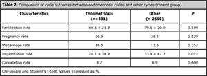
Table 2. Comparison of cycle outcomes between endometriosis cycles and other cycles (control group)

Table 3. Comparison of the presence of oocyte defects between the endometriosis and control groups

Table 4. Comparison of cleavage stage embryo quality and rate of blastocyst formation between the endometriosis and control groups
DISCUSSION
The present study looked into the effects of endometriosis-associated infertility on ICSI outcomes. Significantly lower response to COS, lower oocyte quality, lower fertilization rates, lower cleavage stage embryo quality, and lower implantation rates were observed in individuals with endometriosis. Additionally, the pregnancy rates seen in subjects with endometriosis-associated infertility were comparable to the rates observed in cases of infertility for other causes.
These findings are in line with recent reports showing that women with endometriosis undergoing ART have a significantly lower oocyte yield (Singh et al., 2014) and lower fertilization rates (Opoien et al., 2012) in comparison with tubal factor infertility. In accordance with our findings, Opoien et al. (2012) also described that in addition to the lower fertilization rate, infertile women with endometriosis have comparable pregnancy rates and live birth rates to women with tubal factor infertility, even after adjusting for confounding factors. In a conflicting finding, Opoien´s study reported similar implantation rates between subjects with and without endometriosis.
A previous meta-analysis concluded that patients with endometriosis-associated infertility undergoing IVF respond with significantly lower levels of all markers of the reproductive process, resulting in a pregnancy rate that is almost half of that of women with other indications for IVF. The authors suggested that endometriosis affects not only the receptivity of the endometrium, but also the development of the oocyte and the embryo (Barnhart et al., 2002). More recently, the presence of severe endometriosis was associated with poor implantation in women undergoing IVF (Harb et al., 2013).
A recently published trial by Filippi et al. (2014), investigated whether the presence of endometrioma affected oocyte developmental competence. The study showed that the presence of ovarian endometrioma does not affect oocyte quality. In the study described above, although the authors reported that oocyte morphology was evaluated using an inverted microscope, these data were not described in the results and, apparently, oocyte developmental competence was measured based on the quality of the embryo at the cleavage stage. In our study, oocyte quality was measured based on the presence of individual intra- and extra-cytoplasmic defects. It was noted that when compared to other causes of infertility, endometriosis patients have a higher incidence of extracytoplasmic oocyte defects.
Mature oocytes with apparently normal cytoplasmic organization may exhibit extra-cytoplasmic defects, such as increased perivitelline space, perivitelline debris and/or fragmentation of the first polar body, which may impair the developmental competence of the oocyte (Xia, 1997).
In the present study, in addition to poor oocyte morphology, the fertilization rate was also significantly lower in endometriosis cases. This finding is in accordance with previous studies, showing that poor oocyte morphology is a major determinant of failed or impaired fertilization (Xia, 1997; Javed & Michael, 2012). In addition, a recent meta-analysis published by our group (Setti et al., 2011) showed that the probability of an oocyte becoming fertilized is significantly reduced when extra-cytoplasmic defects such as a large polar body and large perivitelline space are present. Conversely, Balaban & Urman (2006) suggested that extra-cytoplasmic oocyte dysmorphisms should be considered merely as a phenotypic deviation resulting from the heterogeneity of the oocytes retrieved. Nevertheless, as described by Rienzi et al. (2010) in a systematic review of the literature on the predictive value of oocyte morphology in IVF, published studies have produced contradicting results and do not entirely support the typical opinion about the features of ‘good’ and ‘bad’ oocyte quality and developmental competence.
Interestingly, even though oocyte quality and embryo morphology at the cleavage stage were decreased in the endometriosis group, the rate of blastocyst formation did not differ between groups. Previous studies failed to demonstrate (Graham et al., 2000) or demonstrated a poor correlation (Guerif et al., 2010) between early embryo morphology and blastocyst developmental competence. Conversely, we recently investigated the factors that may contribute to the rate of blastocyst formation and quality, and the data showed that good morphology on day three increased the likelihood by up to three-fold that a blastocyst would be of good quality (Braga et al., 2012).
It could be argued that although the presence of endometriosis may have a detrimental effect on oocyte quality and embryo morphology, after prolonged culture to the blastocyst stage, the embryonic genome has begun to be expressed (Tesarik, 2005). At this stage, sperm-derived genes that influence embryo viability have also been activated.
Our results also showed that implantation – but not pregnancy – rates were also decreased in women with endometriosis undergoing ICSI cycles. Whereas some studies (Bukulmez et al., 2001) fail to show decreased implantation rates in IVF patients with endometriosis, others (Kuivasaari et al., 2005) demonstrate significantly lower implantation rates in individuals with early and late stages of the disease.
Embryo implantation depends on three critical events: proper embryo development, the acquisition of a receptive endometrium, and proper dialogue between them (Dominguez et al., 2003). Previous reports have discussed a possible role for endometrial receptivity accounting for lower implantation rates in these patients (Rajani et al., 2012), including decreased expression of implantation markers during the window of implantation (Donaghay & Lessey, 2007; Matsuzaki et al., 2010).
The reason why diminished implantation – but not pregnancy – rates were observed in the present trial is unclear. It may be suggested that the pregnancy rate does not consider the number of transferred embryos, which also differed between groups.
In conclusion, this study showed that in addition to a lower number of oocytes, endometriosis patients also had lower implantation rates. The poor implantation rates observed among endometriosis patients may be explained by the reduced quality of their oocytes and embryos, impaired endometrial receptivity, or perhaps both factors combined.
REFERENCES
Aboulghar MA, Mansour RT, Serour GI, Al-Inany HG, Aboulghar MM. The outcome of in vitro fertilization in advanced endometriosis with previous surgery: a case-controlled study. Am J Obstet Gynecol. 2003;188: 371-5.
Medline Crossref
Al-Azemi M, Bernal AL, Steele J, Gramsbergen I, Barlow D, Kennedy S. Ovarian response to repeated controlled stimulation in in-vitro fertilization cycles in patients with ovarian endometriosis. Hum Reprod. 2000; 15: 72-5.
Medline Crossref
Al-Fadhli R, Kelly SM, Tulandi T, Tanr SL. Effects of different stages of endometriosis on the outcome of in vitro fertilization. J Obstet Gynaecol Can. 2006; 28: 888-91.
Medline
Arici A, Oral E, Bukulmez O, Duleba A, Olive DL, Jones EE. The effect of endometriosis on implantation: results from the Yale University in vitro fertilization and embryo transfer program. Fertil Steril. 1996; 65:603-7.
Medline
Balaban B, Urman B. Effect of oocyte morphology on embryo development and implantation. Reprod Biomed Online. 2006; 12: 608-15.
Medline Crossref
Barnhart K, Dunsmoor-Su R, Coutifaris C. Effect of endometriosis on in vitro fertilization. Fertil Steril. 2002; 77: 1148-55.
Medline Crossref
Braga DP, Setti AS, de Cassia SFR, Machado RB, Iaconelli A Jr, Borges E Jr. Patient selection criteria for blastocyst transfers in extended embryo culture programs. J Assist Reprod Genet. 2012; 29:1357-62.
Medline Crossref
Brizek CL, Schlaff S, Pellegrini VA, Frank JB, Worrilow KC. Increased incidence of aberrant morphological phenotypes in human embryogenesis--an association with endometriosis. J Assist Reprod Genet. 1995, 12: 106-12.
Medline Crossref
Bukulmez O, Yarali H, Gurgan T. The presence and extent of endometriosis do not effect clinical pregnancy and implantation rates in patients undergoing intracytoplasmic sperm injection. Eur J Obstet Gynecol Reprod Biol. 2001; 96:102-7.
Medline Crossref
Burns WN, Schenken RS. Pathophysiology of endometriosis-associated infertility. Clin Obstet Gynecol. 1999; 42: 586-610.
Medline
Chan RW, Schwab KE, Gargett CE. Clonogenicity of human endometrial epithelial and stromal cells. Biol Reprod. 2004; 70: 1738-50.
Medline Crossref
Dominguez F, Pellicer A, Simon C. The chemokine connection: hormonal and embryonic regulation at the human maternal-embryonic interface--a review. Placenta 2003; 24: S48-55.
Medline Crossref
Donaghay M, Lessey BA. Uterine receptivity: alterations associated with benign gynecological disease. Semin Reprod Med. 2007; 25: 461-75.
Medline Crossref
Dong X, Liao X, Wang R, Zhang H. The impact of endometriosis on IVF/ICSI outcomes. Int J Clin Exp Pathol. 2013; 6: 1911-8.
Medline
Du H, Taylor HS. Contribution of bone marrow-derived stem cells to endometrium and endometriosis. Stem Cells. 2007; 25: 2082-6.
Medline Crossref
Filippi F, Benaglia L, Paffoni A, Restelli L, Vercellini P, Somigliana E, Fedele L. Ovarian endometriomas and oocyte quality: insights from in vitro fertilization cycles. Fertil Steril. 2014; 101: 988-93.e1.
Medline Crossref
Garrido N, Navarro J, Remohi J, Simon C, Pellicer A. Follicular hormonal environment and embryo quality in women with endometriosis. Hum Reprod Update. 2000; 6: 67-74.
Medline Crossref
Gianaroli L, Magli MC, Cavallini G, Crippa A, Capoti A, Resta S, Robles F, Ferraretti AP. Predicting aneuploidy in human oocytes: key factors which affect the meiotic process. Hum Reprod. 2010; 25: 2374-86.
Medline Crossref
Giudice LC, Kao LC. Endometriosis. Lancet. 2004; 364:1789-99.
Medline Crossref
Goud PT, Goud AP, Joshi N, Puscheck E, Diamond MP, Abu-Soud HM. Dynamics of nitric oxide, altered follicular microenvironment, and oocyte quality in women with endometriosis. Fertil Steril. 2014; 102: 151-9 e5.
Medline Crossref
Graham J, Han T, Porter R, Levy M, Stillman R, Tucker MJ. Day 3 morphology is a poor predictor of blastocyst quality in extended culture. Fertil Steril. 2000;74: 495-7.
Medline Crossref
Guerif F, Lemseffer M, Leger J, Bidault R, Cadoret V, Chavez C, Gasnier O, Saussereau MH, Royere D. Does early morphology provide additional selection power to blastocyst selection for transfer? Reprod Biomed Online. 2010; 21: 510-9.
Medline Crossref
Gupta S, Agarwal A, Agarwal R, Loret de Mola JR. Impact of ovarian endometrioma on assisted reproduction outcomes. Reprod Biomed Online. 2006; 13: 349-60.
Medline Crossref
Harb HM, Gallos ID, Chu J, Harb M, Coomarasamy A. The effect of endometriosis on in vitro fertilisation outcome: a systematic review and meta-analysis. Bjog. 2013; 120: 1308-20.
Medline Crossref
Holoch KJ, Lessey BA. Endometriosis and infertility. Clin Obstet Gynecol. 2010; 53: 429-38.
Medline Crossref
Inoue M, Kobayashi Y, Honda I, Awaji H, Fujii A. The impact of endometriosis on the reproductive outcome of infertile patients. Am J Obstet Gynecol. 1992; 167: 278-82.
Medline Crossref
Javed M, Michael E. Intracytoplasmic Sperm Injection – Factors Affecting Fertilization. Enhancing Success of Assisted Reproduction. In Darwish AMM (ed) Rijeka: InTech; 2012. p 117-44.
Crossref
Kuivasaari P, Hippelainen M, Anttila M, Heinonen S. Effect of endometriosis on IVF/ICSI outcome: stage III/IV endometriosis worsens cumulative pregnancy and live-born rates. Hum Reprod. 2005; 20: 3130-5.
Medline Crossref
Lin XN, Wei ML, Tong XM, Xu WH, Zhou F, Huang QX, Wen GF, Zhang SY. Outcome of in vitro fertilization in endometriosis-associated infertility: a 5-year database cohort study. Chin Med J (Engl). 2010; 125: 2688-93.
Medline Crossref
Macer ML, Taylor HS. Endometriosis and infertility: a review of the pathogenesis and treatment of endometriosis-associated infertility. Obstet Gynecol Clin North Am. 2012; 39: 535-49.
Medline Crossref
Matalliotakis IM, Cakmak H, Mahutte N, Fragouli Y, Arici A. Sakkas D. Women with advanced-stage endometriosis and previous surgery respond less well to gonadotropin stimulation, but have similar IVF implantation and delivery rates compared with women with tubal factor infertility. Fertil Steril. 2007; 88: 1568-72.
Medline Crossref
Matson PL, Yovich JL. The treatment of infertility associated with endometriosis by in vitro fertilization. Fertil Steril. 1986; 46: 432-4.
Medline
Matsuzaki S, Darcha C, Maleysson E, Canis M, Mage G. Impaired down-regulation of E-cadherin and beta-catenin protein expression in endometrial epithelial cells in the mid-secretory endometrium of infertile patients with endometriosis. J Clin Endocrinol Metab. 2010; 95:3437-45.
Medline Crossref
Meden-Vrtovec H, Tomazevic T, Verdenik I. Infertility treatment by in vitro fertilization in patients with minimal or mild endometriosis. Clin Exp Obstet Gynecol. 2000; 27: 191-3.
Medline
Olive DL, Pritts EA. Treatment of endometriosis. N Engl J Med. 2001; 345: 266-75.
Medline Crossref
Olivennes F, Feldberg D, Liu HC, Cohen J, Moy F, Rosenwaks Z. Endometriosis: a stage by stage analysis--the role of in vitro fertilization. Fertil Steril. 1995; 64: 392-8.
Medline
Opoien HK, Fedorcsak P, Omland AK, Abyholm T, Bjercke S, Ertzeid G, Oldereid N, Mellembakken JR, Tanbo T. In vitro fertilization is a successful treatment in endometriosis-associated infertility. Fertil Steril. 2012; 97: 912-8.
Medline Crossref
Ota H, Igarashi S, Kato N, Tanaka T. Aberrant expression of glutathione peroxidase in eutopic and ectopic endometrium in endometriosis and adenomyosis. Fertil Steril. 2000; 74: 313-8.
Medline Crossref
Ota H, Igarashi S, Sato N, Tanaka H, Tanaka T. Involvement of catalase in the endometrium of patients with endometriosis and adenomyosis. Fertil Steril. 2002; 78: 804-9.
Medline Crossref
Pellicer A, Oliveira N, Ruiz A, Remohi J, Simon C. Exploring the mechanism(s) of endometriosis-related infertility: an analysis of embryo development and implantation in assisted reproduction. Hum Reprod. 1995; 10: 91-7.
Medline Crossref
Rienzi L, Vajta G, Ubaldi F. Predictive value of oocyte morphology in human IVF: a systematic review of the literature. Hum Reprod Update. 2010; 17: 34-45.
Medline Crossref
Setti AS, Figueira RC, Braga DP, Colturato SS, Iaconelli A Jr, Borges E Jr. Relationship between oocyte abnormal morphology and intracytoplasmic sperm injection outcomes: a meta-analysis. Eur J Obstet Gynecol Reprod Biol. 2011; 159: 364-70.
Medline Crossref
Simon C, Gutierrez A, Vidal A, de los Santos MJ, Tarin JJ, Remohi J, Pellicer A. Outcome of patients with endometriosis in assisted reproduction: results from in-vitro fertilization and oocyte donation. Hum Reprod. 1994; 9: 725-9.
Medline
Singh N, Lata K, Naha M, Malhotra N, Tiwari A, Vanamail P. Effect of endometriosis on implantation rates when compared to tubal factor in fresh non donor in vitro fertilization cycles. J Hum Reprod Sci. 2014; 7: 143-7.
Medline Crossref
Steele RW, Dmowski WP, Marmer DJ. Immunologic aspects of human endometriosis. Am J Reprod Immunol. 1984; 6: 33-6.
Medline Crossref
Tanbo T, Omland A, Dale PO, Abyholm T. In vitro fertilization/embryo transfer in unexplained infertility and minimal peritoneal endometriosis. Acta Obstet Gynecol Scand. 1995; 74: 539-43.
Medline Crossref
Tesarik J. Paternal effects on cell division in the human preimplantation embryo. Reprod Biomed Online. 2005; 10: 370-5.
Medline Crossref
Vercellini P, Vigano P, Somigliana E, Fedele L. Endometriosis: pathogenesis and treatment. Nat Rev Endocrinol. 2013; 10: 261-75.
Medline Crossref
Verkauf BS. Incidence, symptoms, and signs of endometriosis in fertile and infertile women. J Fla Med Assoc. 1987; 74: 671-5.
Medline
Wardle PG, Mitchell, JD, McLaughlin EA, Ray BD, McDermott A, Hull MG. Endometriosis and ovulatory disorder: reduced fertilisation in vitro compared with tubal and unexplained infertility. Lancet. 1985; 2: 236-9.
Medline Crossref
Xia P. Intracytoplasmic sperm injection: correlation of oocyte grade based on polar body, perivitelline space and cytoplasmic inclusions with fertilization rate and embryo quality. Hum Reprod. 1997; 12: 1750-5.
Medline Crossref
Zanatta A, Rocha AM, Carvalho FM, Pereira RM, Taylor HS, Motta EL, Baracat EC, Serafini PC. The role of the Hoxa10/HOXA10 gene in the etiology of endometriosis and its related infertility: a review. J Assist Reprod Genet. 2010; 2712: 701-10.
Medline Crossref