JBRA Assist. Reprod. 2016;20 (1):44-46
CASE REPORT
doi: 10.5935/1518-0557.20160011
A successful healthy live birth from a female patient with hypogonadotropic hypogonadism and oocytes with unusually large cytoplasmic inclusions
1Turgut Ozal University, School of Medicine, Department of Obstetrics and Gynecology, IVF Clinic, Ankara, Turkey
CONFLICT OF INTERESTS
No conflict of interest have been declared.
ABSTRACT
This study aimed to report the case of a successful live birth from a
woman having oocytes with abnormally large cytoplasmic inclusions. The
patient described in this case is a 28 year-old woman with
hypogonadotropic hypogonadism (HH) with a history of two previous
unsuccessful in vitro fertilization (IVF) attempts offered an
antagonist protocol. Stimulation was performed with human menopausal
gonadotropin 300 IU/day. The intracytoplasmic sperm injection (ICSI)
procedure was performed 4-6 hours after oocyte aspiration for all
mature oocytes. Six oocytes were retrieved, five of which mature (MII).
All oocytes had abnormal cytoplasmic structures. Two were fertilized
after ICSI and two top quality embryos were transferred on Day 2. Our
case report suggests that HH patients with refractile bodies/lipofuscin
in their oocytes may not have their pregnancies negatively affected.
While there have been several reports of successful births from
dysmorphic oocytes, no cases of successful pregnancies followed by live
births from young women with HH and oocytes with large cytoplasmic
inclusions had been reported to date.
Keywords: Refractile body, Lipofuscin, Oocyte Cytoplasm, ICSI
INTRODUCTION
Oocyte morphology is an important factor in female fertility and a
reflection of the intrinsic developmental potential of the oocyte;
additionally, it plays an important role in fertilization and
subsequent embryo development (Gilchrist et al., 2008).
According to some data, the phenotype of the adult stage offspring is
considerably defined by the quality of the oocytes from which they are
derived (Mtango et al., 2008). Different
kinds of oocyte dysmorphisms have been identified in humans. They can
be extracytoplasmic or cytoplasmic anomalies (Mikkelsen & Lindenberg, 2001; Ebner et al., 2006).
It has been reported that the development of intracytoplasmic and
extracytoplasmic anomalies during the maturation process may lead to
fertilization failure (Ebner et al., 2006; De Sutter et al., 1996; Rienzi L et al., 2008), chromosome aneuploidy (Kahraman et al., 2000; Van Blerkom & Henry,1992), and developmental impairment of the embryo despite normal fertilization (Xia P, 1997).
Analysis of the literature, however, shows that the effect of oocyte
morphologic deviations after ICSI remains controversial (Balaban & Urman, 2006; Balaban et al., 1998). In the literature, different anomalies of oocyte morphology have been described (Van Blerkom & Henry,1992).
Refractile bodies are one of the main morphological abnormalities in
the cytoplasm of oocytes. They include lipid bodies, small autophagic
vacuoles, and lipofuscin bodies. These inclusions may range in size
from 1 to 5 µm. Large inclusions (> 5 µm) have been associated with
lower chance of fertilization and blastocyst development rate (Otsuki et al., 2007).
In 1912, Hueck presented the term lipofuscin, based on lipo (Greek for
fat) and fuscus (Latin for dark), consisting of aggregated polymers
derived from the oxidation products of lipids and proteins. Lipofuscin
is autofluorescent (Browne & Rippin, 1977; Collins & Brunk, 1976) and related to aging with continuous accumulation over time within post-mitotic cells such as neurons (Glees & Hasan, 1976; Sohal & Brunk, 1989) and cardiac myocytes (Munnell & Getty, 1968).
Experimental data have shown that the cytoplasmic accumulation of
lipofuscin in cells is accelerated by increased oxygen levels (Sohal & Brunk, 1989; Munnell & Getty, 1968; Gao, 1994 ) and by inhibitors of lysosomal proteases and lipases (Marzabadi et al., 1991),
suggesting that oxidative stress and the incomplete degradation of
autophagocytosed material may cause lipofuscinogenesis by the
accumulation of peroxidized lipids and proteins in secondary lysosomes (Terman, 1995).
The underlying mechanism that causes the occurrence of refractile
bodies is unknown. A correlation between morphology, developmental
competence, and chromosomal abnormalities has not been clearly
established.
CASE REPORT
A 28-year old woman
diagnosed with hypogonadotropic hypogonadism with a history of two
previous unsuccessful IVF attempts came to our IVF clinic in March of
2013. Her hormone profile on the third day of menstruation cycle was as
follows: FSH: 0.6 mIU/ml, E2: 22.7 pg/ml, LH: <0.07 mIU/ml, PRL: 9.4
ng/ml, P2: 0.3 ng/ml, AMH: 0.3 ng/ml. Controlled ovarian stimulation
was achieved with the use of human menopausal gonadotropin (HMG) 375
IU/day as a daily dose, starting on Day 3 of the cycle. Follicular
development was monitored by serial transvaginal ultrasonography and
serum E2 levels. Pituitary blockage was performed with a GnRH
antagonist (Cetrotide; Serono) given when at least one follicle reach
14 mm. When the mean follicle size reached 17 mm, an injection of 250
µg of recombinant human chorionic gonadotropin (hCG, Ovitrelle; Serono)
was given to trigger the final stages of oocyte maturation;
ultrasound-guided oocyte pick-up was performed 35-36 hours later. Six
oocytes were retrieved, five of which mature (MII). The
intracytoplasmic sperm injection (ICSI) procedure was performed 4-6
hours after oocyte aspiration for all mature oocytes. Fertilization
check was done 16-18 hours after ICSI. The embryos were transferred on
Day 2. The luteal phase was supported with a daily 8% progesterone gel
(Crinone; Serono) started on the day of oocyte retrieval (Table 1).
Two of the mature oocytes were fertilized and cleaved in culture
medium. Two 4-cell grade I embryos were transferred on Day 2. A
pregnancy test carried out 12 days after transfer showed a β-hCG level
of 62.7 mIU/mL, which increased to 158 mIU/mL two days later. A
singleton pregnancy with a visible heart beat was detected at
gestational week 7. All oocytes had abnormal cytoplasmic structures,
resembling an accumulation of refractile bodies, although atypically
large (Figure1).
However, the material could be the same, which is lipofuscin. They did
not disappear after fertilization, and were still seen after cleavage
in one blastomere (Figure 2).
According to the literature, trace inclusions may persist up to the
blastocyst stage. However, larger inclusions usually end up in a cell
which either stops developing or goes to trophectoderm. But since this
inclusion was too big for a trophectoderm cell, the affected cell may
be excluded from the embryo.
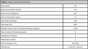
Table 1. Patient clinical characteristics
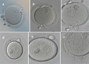
Figure 1: Large fractile body/lipofuscin in the oocytes (a,b,c,d,e,f)
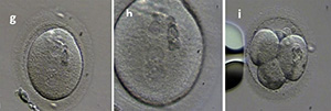
Figure 2: Large refractile body/lipofuscin after fertilization (g,h) and in one blastomere of embryos (i)
DISCUSSION
The female gamete plays a crucial role in determining embryo competence
and, therefore, in vitro fertilization (IVF) outcomes. Oocyte quality
is influenced by the nuclear and mitochondrial genome and by the
microenvironment provided by the ovary and the pre-ovulatory follicle,
which by its turn affects transcription and translation and,
consequently, cytoplasmic maturity. In contrast with in vivo processes,
the use of ovarian stimulation in human reproduction further
complicates the already complex selection procedure that usually occurs
during oocyte development and maturation of a single oocyte for
ovulation, while allowing for the maturation of many oocytes, often
with compromised quality (Rienzi et al., 2008).
It has been shown in the literature that severe dysmorphisms of the
cytoplasmic texture impairs the developmental and implantation
potential of the embryo (Balaban & Urman, 2006).
According to some authors, cytoplasmic inclusions do not seem to affect
fertilization, embryo quality, or implantation rates (De Sutter et al., 1996; Balaban & Urman, 2006). Others, however, have reported decreased fertilization and embryo development (Xia, 1997; Otsuki et al., 2007).
Negative impacts from specific intracytoplasmic and extracytoplasmic
oocyte defects on the blastocyst quality parameters have been reported (Braga et al., 2013).
Hypogonadotropic hypogonadism (HH) has been classified as a Group I
anovulation disorder by the World Health Organization (WHO).
Individuals with HH are good candidates for ovulation induction with
exogenous gonadotropins. HH is usually idiopathic, with no anatomical
lesions in the pituitary stalk.
The condition is characterized by amenorrhea, hypoestrogenism, low
serum gonadotropins, and a broad spectrum of abnormal secretion
patterns of hypothalamic gonadotropin releasing hormone (GnRH) (Reame et al., 1985).
Ovulation induction can be achieved with small doses of menotropins.
The conventional procedure is to start ovulation with lower daily doses
of human menopausal gonadotropins (hMG). In our patient the starting
dose of menotropins was high and the course of gonadotropin
administration was long (12 days). The high doses prescribed to the
patient may have affected oocyte metabolism. The occurrence of large
refractile bodies/lipofuscin bodies may be related to the status of
perifollicular blood circulation and follicular fluid composition seen
in patients with HH.
There are no studies comparing abnormal oocyte
morphology according to infertility etiology. The formation of
refractile bodies/lipofuscin has been correlated to oxidative stress (Veeck, 1991).
It has also been suggested that the occurrence of lipofuscin may be
related to the metabolism of lipids into energy. Potential interactions
between adipokines and the hypothalamus, pituitary gland, ovary,
oocyte, embryo, and female reproductive tract such as insulin
resistance have been suggested (Van Blerkom, 1990).
CONCLUSIONS
The predictive value of oocyte cytoplasmic morphology has been a topic
of controversy in the literature. Our case report suggests that the
pregnancies of patients with HH and refractile body/lipofuscin in their
oocytes may not be adversely affected. While there have been several
reports of successful births from dysmorphic oocytes, no cases of
successful pregnancies followed by live births from women with HH and
oocytes with large cytoplasmic inclusions had been reported to date.
REFERENCES
Balaban
B, Urman B, Sertac A, Alatas C, Aksoy S, Mercan R. Oocyte morphology
does not affect fertilization rate, embryo quality and implantation
rate after intracytoplasmic sperm injection. Hum Reprod 1998;13:3431–3.
Medline Crossref
Balaban B, Urman B. Effect of oocyte morphology on embryo development and implantation Reprod Biomed Online. 2006;12:608-15.
Medline Crossref
Braga
DP, Setti AS, Figueira Rde C, Machado RB, Iaconelli A Jr, Borges E Jr.
Influence of oocyte dysmorphisms on blastocyst formation and quality.
Fertil Steril. 2013;100:748-54.
Medline Crossref
Browne RM, Rippin JW. Autofluorescent granular cells in oral mucosal hyperplasias. Histopathology. 1977;1:375–84.
Medline Crossref
Collins
VP, Brunk UT. Characterization of residual bodies formed in phase II
cultivated human glia cells. Mech Ageing Dev 1976;5:193–207.
Medline Crossref
De
Sutter P, Dozortsev D, Qian C, Dhont M. Oocyte morphology does not
correlate with fertilization rate and embryo quality after
intracytoplasmic sperm injection. Hum Reprod 1996;11:595–7.
Medline Crossref
Ebner
T, Moser M, Tews G. Is oocyte morphology prognostic of embryo
developmental potential after ICSI? Reprod Biomed Online. 2006; 12:
507–12.
Medline Crossref
Gao
G, Ollinger K, Brunk UT. Influence of intracellular glutathione
concentration of lipofuscin accumulation in cultured neonatal rat
cardiac myocytes. Free Radic Biol Med 1994;16:187–94.
Medline Crossref
Gilchrist
RB, Lane M, Thompson JG. Oocyte-secreted factors: regulators of cumulus
Cell function and oocyte quality. Hum Reprod Update 2008;14:159–77.
Medline Crossref
Glees P, Hasan M. Lipofuscin in neuronal aging and diseases. Norm Pathol Anat 1976; 32:1–68.
Medline
Kahraman
S, Yakin K, Donmez E, Samli H, Bahce M, Cengiz G, Sertyel S, Samli M,
Imirzalioğlu N. Relationship between granular cytoplasm of oocytes and
pregnancy outcome following intracytoplasmic sperm injection. Hum
Reprod. 2000;15:2390–3.
Medline Crossref
Marzabadi
MR, Sohal RS, Brunk UT. Mechanisms of lipofuscinogenesis: effect of the
inhibition of lysosomal proteinases and lipases under varying
concentrations of ambient oxygen in cultured rat neonatal myocardial
cells. APMIS. 1991; 99:416–26.
Medline Crossref
Mikkelsen
AL, Lindenberg S. Morphology of in-vitro matured oocytes: impact on
fertility potential and embryo quality. Hum Reprod. 2001; 16: 1714–8.
Medline Crossref
Mtango NR, Potireddy S, Latham K. Oocyte quality and maternal control of development. Int Rev Cell Mol Biol 2008;268:223–90.
Medline Crossref
Munnell JF, Getty R. Rate of accumulation of cardiac lipofuscin in the aging canine. J Gerontol 1968;23:154–8.
Medline Crossref
Otsuki
J, Nagai Y, Chiba K. Lipofuscin bodies in human oocytes as an indicator
of oocyte quality. J Assist Reprod Genet 2007;24:263–70.
Medline Crossref
Reame
NE, Sauder SE, Case GD, Kelch RP, Marshall JC. Pulsatile gonadotropin
secretion in women with hypothalamic amenorrhea: Evidence that reduced
frequency of gonadotropin-releasing hormone secretion is the mechanism
of persistent anovulation. J Clin Endocrinol Metab 1985;61:851–858.
Medline Crossref
Rienzi
L, Ubaldi FM, Iacobelli M, Minasi MG, Romano S, Ferrero S, Sapienza F,
Baroni E, Litwicka K, Greco E. Significance of metaphase II human
oocyte morphology on ICSI outcome. Fertil Steril 2008;90:1692–700.
Medline Crossref
Sohal RS, Brunk UT. Lipofuscin as an indicator of oxidative stress and aging. Adv Exp Med Biol 1989;266:17–29.
Medline
Terman
A. The effect of age on formation and elimination of autophagic
vacuoles in mouse hepatocytes. Gerontology 1995;41:319–26.
Medline Crossref
Van
Blerkom J. Occurrence and developmental consequences of aberrant
cellular organization in meiotically mature human oocytes after
exogenous ovarian hyperstimulation. J ElectronMicroscTech
1990;16:324–46.
Medline Crossref
Van
Blerkom J, Henry G. Oocyte dysmorphism and aneuploidy in meiotically
mature human oocytes after ovarian stimulation. Hum Reprod 1992;7:
379–90.
Medline
Veeck LL, ed. Atlas of the human oocyte and early conceptus. Baltimore: Williams & Wilkins; 1991.
Xia
P. Intracytoplasmic sperm injection: correlation of oocyte grade based
on polar body, perivitelline space and cytoplasmic inclusions with
fertilization rate and embryo quality. Hum Reprod 1997;12:1750–5.
Medline Crossref