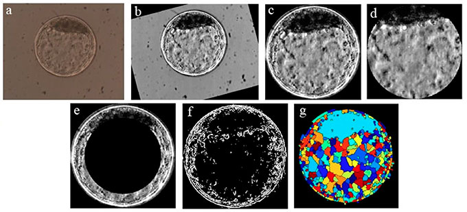
Figure 1. Ilustrates the sequence of steps required to process a digital image from an in vitro produced bovine blastocyst. a) original image as obtained by optic microscopy; b) standardization of bright and positioning of the inner cell mass (ICM) at 12 o’clock; c) segmentation of the embryo itself (by Hough transform) and elimination of background; d) segmentation of ICM and blastocoel by elimination of the zona pellucida and trophectoderm; e) elimination of inner area of the image “c” to highlight the trophectoderm and part of the ICM; f) binary form of image “c” after gradient calculation; g) visualization of the image after Watershed transform.