JBRA Assist. Reprod. 2016;20 (4):253-256
CASE REPORT
doi: 10.5935/1518-0557.20160049
Live births after polar body biopsy and frozen-thawed cleavage stage embryo transfer: case report
1ORIGEN – Center for Reproductive Medicine, Rio de Janeiro/RJ - Brazil
2UFMG – Universidade Federal de Minas Gerais, Belo Horizonte/MG - Brazil
3Chromosome Genomic Medicine, São Paulo/SP - Brazil
4ORIGEN – Center for Reproductive Medicine, Belo Horizonte/MG - Brazil
CONFLICT OF INTERESTS
No conflict of interest have been declared.
ABSTRACT
Pre-implantation genetic diagnosis (PGD) or screening (PGS) technology, has emerged and developed in the past few years, benefiting couples as it allows the selection and transfer of healthy embryos during IVF treatments. These techniques can be performed in oocytes (polar-body biopsy) or embryos (blastomere or trophectoderm biopsy). In this case report, we describe the first two live births to be published in Brazil after a polar-body (PB) biopsy. In case 1, a 42-year-old was submitted to PB biopsy with PGS due to advanced maternal age and poor ovarian reserve. Five MII oocytes underwent first and second polar body biopsy and four cleavage embryos were cryopreserved. The PGS analysis resulted in two euploid embryos (next generation sequence). A frozen-thawed embryo transfer (FET) was performed after endometrial priming and a healthy baby was delivered after a cesarean section (37 weeks, female, 3390g, 47.5 cm). In case 2, a 40-year old patient with balanced translocation and poor ovarian response was submitted to PB biopsy. Two MII oocytes underwent first and second polar body biopsy and two embryos were cryopreserved in cleavage stage. The analysis resulted in one euploid embryo that was transferred after endometrial priming. A preterm healthy baby (34 weeks, female, 2100g, 40 cm) was delivered via cesarean section. In conclusion, although the blastocyst biopsy is the norm when performing PGS/PGD during IVF treatments, other alternatives (as PB biopsy) should be considered in some specific situations.
Keywords: Polar body biopsy, pre-implantation genetic screening, pre-implantation genetic diagnosis, PGS, PGD.
INTRODUCTION
In recent years, the development of assisted reproduction techniques (ART) has provided enormous advances to the treatment of infertile couples. One of the areas of development has been pre-implantation genetic analysis, with the use of preimplantation genetic diagnosis (PGD) or screening (PGS) technology. These techniques have benefited healthy couples and also those with a history of genetic disorders, enabling the selection and transfer of healthy embryos (Harper, 2009).
Chromosome aneuploidy is one of the major concerns related to advanced maternal age in women seeking pregnancy. It is associated with pregnancy failure, miscarriage and chromosomal anomalies in the offspring after natural or assisted conceptions (Christopikou et al., 2013). Pre-implantation genetic evaluation is a powerful tool to achieve a healthy pregnancy, avoiding potential adverse events related to advanced maternal age and chromosomal anomalies (Cimadomo et al., 2016).
Pre-implantation analysis can be performed in three developmental stages: oocyte (polar body biopsy); cleavage stage (blastomere biopsy); and finally: blastocyst stage (trophectoderm biopsy) (Kanavakis & Traeger-Synodinos, 2002). To perform the biopsy, it is important to cause a disruption of the zona pellucida of the oocyte or embryo occurs, which can be performed mechanically, chemically or using laser (Brezina et al., 2012). The key point of PGD/PGS is to have access to the genetic material to be evaluated, without compromising the material analyzed and the quality of the oocyte/embryo (Xu & Montag, 2012).
Polar body (PB) biopsy was introduced in 1990 (Verlinsky et al., 1990), and it is associated with a less invasive technique, presenting advantages, because it maintains embryo integrity, as only meiotic products are used to assess oocyte conditions. Removal of the first and/or second polar body is an indirect approach to evaluate oocyte genetic or chromosomal status (Montag et al., 2013). Moreover, the use of polar body biopsy is an option for countries with strict regulations regarding embryo biopsy, as it is the case in Germany and Switzerland (Harton et al., 2011; Xu & Montag, 2012). A pilot study (ESHRE PGS Task force) for PGS in women of advanced maternal age was conducted. Polar bodies following intracytoplasmic sperm injection (ICSI) were evaluated by CGH array to detect copy number changes and to predict aneuploidy status in the corresponding embryos. This study demonstrated that polar body and CGH array analysis were efficient and was highly concordant (94%) with the statuses of the corresponding zygotes (Christopikou et al., 2013). A recent publication from 2011 reviewed 938 cycles of PGD for 146 types of Mendelian monogenic diseases. They performed 9036 PB biopsies which were evaluated by multiplex PCR, with 1578 healthy embryos, which were later transferred. That resulted in 329 pregnancies and 345 births. This data, demonstrated an important conclusion regarding the safety and efficacy of the use of PGD with PB biopsy (Kuliev & Rechitsky, 2011). Nevertheless, it is hampered by the impossibility of diagnosing paternally derived defects, and those originating after fertilization or first cleavage events (Gianarolli L, 2000).
The main objective of this study was to describe the live birth of two healthy babies in Brazil, after performing a PB biopsy.
CASE DESCRIPTION
Case 1
A 42-year-old patient was referred to our center in November 2014 for an infertility workup. The patient provided a signed informed consent. The couple has a 7 –year-old son with autism. There was no male factor associated. She had a regular menstrual cycle and altered basal hormonal levels, as measured on day 2: follicle-stimulating hormone (FSH), 16 mIU/mL; luteinizing hormone (LH), 6,1 IU/mL; E2, 65 pg/mL. The antral follicle count (AFC) performed on day 3 showed 6 antral follicles.
Ovulation induction started on day 2 of her menstrual cycle in a step-down GnRH antagonist protocol, with 300 IU of recombinant FSH (rFSH; Gonal-F; Merck, Darmstadt, Germany) and 150 IU rFSH with 75 IU rLH (Pergoveris Merck SA, Aubonne, Swiss). The GnRH antagonist (Cetrotide; Merck) was introduced from day 5 of stimulation. Final oocyte maturation was induced when three follicles reached 18 mm in diameter, after 10 days of stimulation, with a bolus of 0.2 mg of GnRHa (triptorelin; Gonapeptyl daily; Ferring; Kiel; Germany). The ultrasound examination on the trigger day showed three follicles ≥ 18 mm, and three follicles between 14–18 mm in diameter. Oocyte retrieval was performed 36 hours after the trigger. A total of five oocytes were retrieved; five were metaphase II (MII), and all of them were inseminated (day 0) by intracytoplasmic sperm injection (ICSI). The first polar body (PB) biopsy was performed at the moment of the ICSI procedure. On the following day of ICSI (day 1), five oocytes were normally fertilized and all oocytes had the second polar body removed. On day 3, there were four cleaved embryos, and both the first and second polar bodies of these embryos were sent for analysis by NGS (next generation sequencing). Fifteen days after, we received the report showing two euploid embryos and two aneuploidy embryos.
Four months later, the patient started endometrial priming with estradiol valerate (6mg/day) on the second day of the cycle. On the 12th day of priming, she had normal hormonal measurements and an ultrasound scan showed an endometrial thickness of 7.5mm. The patient was started on vaginal progesterone. On the fourth day of progesterone administration, two euploid embryos (Fig. 1) were thawed and transferred on the same day. The patient became pregnant. In the 37th week of pregnancy, after a cesarean section, a healthy baby (female, 3390g; 47.5cm) was delivered. There were no obstetrical / perinatal complications.
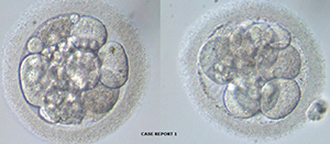
Figure 1. Two euploid embryos transferred - patient 1
Case 2
A 40-year-old patient with balanced translocation was referred to our center. The patient provided a signed informed consent. There was no male factor associated. She had a regular menstrual cycle and normal basal hormonal levels, as measured on day 2: follicle-stimulating hormone (FSH) of 11,2 mIU/mL; luteinizing hormone (LH) of 5,6 IU/mL, and E2 of 49 pg/mL. The basal ultrasound showed four antral follicles. The karyotype showed a balanced translocation - 45XX rob(13.14)q10. Her partner had a normal karyotype.
Ovulation induction started on day 2 of her menstrual cycle in a step-down GnRH antagonist protocol, with 300 IU of recombinant FSH (rFSH; Gonal-F; Merck, Darmstadt, Germany) and 150 recombinant FSH with recombinant LH (Pergoveris Merck SA, Aubonne, Swiss). The GnRH antagonist (Cetrotide; Merck) was introduced as of day 5 of stimulation. Final oocyte maturation was induced when three follicles reached 18 mm in diameter, after 10 days of stimulation, with a bolus of 0.2 mg of GnRHa (triptorelin; Gonapeptyl daily; Ferring; Kiel; Germany). The ultrasound scan showed three follicles ≥ 18 mm in diameter, and two follicles between 16–18 mm in diameter. Oocyte retrieval was performed 36 hours after the trigger. A total of three oocytes were retrieved and two of them were MII. The mature oocytes were inseminated (day 0) by intracytoplasmic sperm injection (ICSI). The first polar body (PB) biopsy was performed at the moment of the ICSI procedure. On the following day of ICSI (day 1), two oocytes were normally fertilized and all oocytes had the second polar body removed. On day 2, there were two cleaved embryos, and both the first and second polar bodies of these embryos were sent for analysis by NGS (next generation sequencing). The NGS analysis showed one euploid embryo and one aneuploid embryo. Two months later, she started endometrial priming with estradiol valerate administration on the second day of the cycle, 6 mg orally daily. On the 12th day of priming she had normal hormonal measurements and an ultrasound scan showed an endometrial thickness of 8mm. Micronized vaginal progesterone (Crinone) was introduced, and on the third day of administration, one euploid embryo (Fig. 2) was thawed and transferred on the same day. The patient became pregnant and delivered a pre-term baby on the 34th week of gestation (female, 2100g; 40cm).
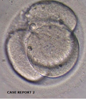
Figure 2. One euploid embryo – patient 2
DISCUSSION
To our knowledge these are the first two live births to be published after PB biopsy in Brazil. The diagnosis of polar body refers to an indirect method of genetically evaluating the oocyte and it seems to be a feasible technique to be employed in some specific situations. This paper presents two patients in which three recent, developing, and highly promising technologies were employed successfully: PB biopsy; cytogenetic evaluation with NGS; and embryo vitrification.
Currently, specific indications for performing PGD/PGS are: patients with advanced maternal age, monogenic diseases, maternal translocations, recurrent implantation failure and recurrent spontaneous abortion (Wang et al., 2010). Although the blastocyst biopsy is the current norm in clinical practice for PGS/PGD, PB biopsy may be an alternative for selected cases. However it is very important that there is an accurate alignment between the clinical evaluation of the patient with the desired type of laboratory technique, as well as proficiency and expertise (Van der Ven et al, 2008).
There are important positive considerations concerning the PB biopsy. First, the occurrence in some countries of stringent regulations or restrictions on the use of biopsy in embryonic stages as Cleavage and Blastocyst, enable only the performance of PB biopsy. Second, this type of procedure is characterized by reduced invasiveness and may not be as harmful as biopsies in more advanced stages, such as Cleavage and Blastocyst (Harton et al; 2011). Third, it may be considered in patients with poor ovarian response, with indication of PGD/PGS. Due to the low number of oocytes in this group of patients, sometimes there are no available embryos to be evaluated in the blastocyst stage. The two reported cases in this manuscript were related to poor ovarian response. All the available strategies were discussed with the patients and the PB biopsy was selected.
It is important to consider some issues and limitations of this technique. The predictive value of aneuploidy is considered impaired when compared blastomere or trophectoderm biopsies, which often require confirmatory analysis. Furthermore, this technique cannot detect genetic errors from the paternal side. Also, when there is a large number of oocytes to be tested, there is a decreased cost-effectiveness of this technique. Another issue that may arise is that polar bodies are very small in size, and eventually the biopsy samples may degenerate or simply not yield results (Xu & Montag, 2011).
Therefore, if PB biopsy will be performed, safe removal of the two polar bodies is mandatory. Thus, both can be analyzed by specific techniques for genetic analysis and a healthy embryo can be selected for embryo transfer (Wei et al., 2015). Important considerations must be taken into account when using this strategy: laboratory techniques, biopsy procedure, the freezing and thawing process, and finally the genetic analysis. Critical steps in this procedure are the opening of the zona pellucida of the oocyte, and the correct and non-traumatic removal of the first and second polar bodies (Magli et al., 2004).
As many embryo transfers are not performed in a fresh cycle when PGD/PGS is carried out, appropriate and validated embryo cryopreservation protocols are essential for achieving good results in this situation, to improve survival and implantation rates (Kuwayama M., 2007; Lee & Munne, 2000). In this study, all euploid embryos survived after the thawing process. In our IVF center, we have achieved good outcomes with embryo cryopreservation with over 90% survival rates when performing vitrification in the cleavage stage (Roque et al., 2015). Moreover, a well-trained staff with experience in embryo biopsy techniques is of great importance to reduce misdiagnoses. The introduction of new technologies in genetic analysis (such as aCGH, SNP microarrays, qPCR and NGS) was paramount in order to achieve the expected clinical benefits that could not be demonstrated by previous techniques such as the FISH analysis. These new methods enable the simultaneous evaluation and identification of embryos with specific chromosomal abnormalities (unbalanced translocations for example) and aneuploidy, thus enabling the selection of euploid embryo for later transfer. However, there is a need for more randomized controlled trials evaluating these new technologies, so as to calculate the real benefits of these strategies, and to pinpoint in which groups of patients it should be performed (Martin et al., 2013).
In conclusion, although blastocyst biopsy is the norm when performing PGS /PGD during IVF treatments; other alternatives, such as PB biopsy, should be taken into account in some specific situations, as in patients with poor ovarian response and a diagnosed female genetic abnormality.
REFERENCES
Brezina PR, Brezina DS, Kearns WG. Preimplantation genetic testing. BMJ 2012; 18:345:e5908.
Medline Crossref
Christopikou D, Tsorva E, Economou K, Shelley P, Davies S, Mastrominas M, Handyside AH. Polar body analysis by array comparative genomic hybridization accurately predicts aneuploidies of maternal meiotic origin in cleavage stage embryos of women of advanced maternal age. Hum Reprod 2013; 28:1426–34.
Medline Crossref
Cimadomo D, Capalbo A, Ubaldi FM, Scarica C, Palagiano A, Canipari R, Rienzi L. The Impact of Biopsy on Human Embryo Developmental Potential during Preimplantation Genetic Diagnosis. BioMed Res Int. 2016;2016:7193075.
Medline Crossref
Gianarolli L. Preimplantation genetic diagnosis: polar body and embryo biopsy. Hum Reprod 2000; 15:69-75.
Medline Crossref
Harper J, ed. Preimplantation Genetic Diagnosis. 2a ed. Cambridge: Cambridge University Press; 2009.
Harton GL, Magli MC, Lundin K, Montag M, Lemmen J, Harper JC. ESHRE PGD Consortium/Embryology Special Interest Group–best practice guidelines for polar body and embryo biopsy for preimplantation genetic diagnosis/screening (PGD/PGS). Hum Reprod 2011; 26: 41–6.
Medline Crossref
Kanavakis E, Traeger-Synodinos J. Preimplanation genetic diagnosis in clinical practice. J Med Genet. 2002; 39: 6-11.
Medline Crossref
Kuliev A, Rechitsky S. Polar body-based preimplantation genetic diagnosis for Mendelian disorders. Mol Hum Reprod. 2011; 17: 275–85.
Medline Crossref
Kuwayama M. Highly efficient vitrification for cryopreservation of human oocytes and embryos : the Cryotop method. Theriogenology 2007; 67:73-80.
Medline Crossref
Lee M, Munne S. Pregnancy after polar body biopsy and freezing and thawing of human embryos. Fertil Steril 2000; 73: 645-7.
Medline Crossref
Magli MC, Gianaroli L, Ferraretti AP, Toschi M, Esposito F, Fasolino MC. The combination of polar body and embryo biopsy does not affect embryo viability. Hum Reprod. 2004; 19: 1163-9.
Medline Crossref
Martin J, Cervero A, Mir P, Martinez-Conejero JA, Pellicer A, Simón C. The impact of next-generation sequencing technology on preimplantation genetic diagnosis and screening. Fertil Steril 2013;99:1054–61.
Medline Crossref
Montag M, Köster M, Strowitzki T, Toth B. Polar body biopsy. Fertil Steril. 2013; 100: 603–7.
Medline Crossref
Roque M, Valle M, Guimarães F, Sampaio M, Geber S. Freeze-all policy: fresh vs. frozen-thawed embryo transfer. Fertil Steril 2015; 103: 1190-3.
Medline Crossref
van der Ven K, Montag M, Van Der Ven H. Polar body diagnosis - a step in the right direction? Dtsch Arztebl Int. 2008; 105:190–6.
Medline Crossref
Verlinsky Y, Ginsberg N, Lifchez A, Valle J, Moise J, Strom CM. Analysis of the first polar body: preconception genetic diagnosis. Hum Reprod 1990; 5: 826–9.
Medline
Wang N, Zheng YM, Li L, Jin F. Preimplantation Genetic Screening: An Effective Testing for Infertile and Repeated Miscarriage Patients? Obstet Gynecol Int. 2010; 2010:120130.
Medline Crossref
Wei Y, Zhang T, Wang YP, Schatten H, Sun QY. Polar bodies in ART: Current Progress and future perspectives . Biol Reprod 2015;92:1-8.
Medline Crossref
Xu K, Montag M. New Perspectives on Embryo Biopsy: Not How, But When and Why? Semin Reprod Med 2012;30:259–66.
Medline Crossref