JBRA Assist. Reprod. 2016;20 (4):200-205
ORIGINAL ARTICLE
doi: 10.5935/1518-0557.20160039
miR-142-3p as a biomarker of blastocyst implantation failure - A pilot study
1Fertility Medical Group – São Paulo/SP - Brazil
2Instituto Sapientiae – Centro de Estudos e Pesquisa em Reprodução Assistida – São Paulo/SP - Brazil
3Disciplina de Urologia, Área de Reprodução Humana, Departamento de Cirurgia, Universidade Federal de São Paulo/SP - Brazil
4Departamento de Biologia Estrutural e Funcional, Instituto de Biologia, Universidade Estadual de Campinas UNICAMP/SP - Brazil
CONFLICT OF INTERESTS
No conflict of interest have been declared.
ABSTRACT
Objective: This study aims to find whether microRNAs (miRNAs) detected in the culture medium of embryos produced in vitro could be potential biomarkers of embryo implantation.
Methods: Culture media samples from 36 embryos, derived from patients undergoing intracytoplasmic sperm injection (ICSI) in a private university-affiliated IVF center, were collected between January/2015 and November/2015. Samples were collected on day three and embryo transfers were performed on day five and all embryos reached the blastocyst stage. Samples were split into groups according to the embryo implantation result: Positive-Implantation-Group (n=18) or Negative-Implantation-Group (n=18). For the first analysis, samples were pooled in three sets for each group (6-7 spent media per pool). MicroRNAs were extracted from spent media and cDNA was synthesized. C. elegans miR-39 was used as RNA spike-in to normalize the gene expression analysis. The expression of microRNAs into the spent media from the Positive-Implantation-Group was compared with those from the Negative-Implantation-Group. A set of seven miRNAs (miR-21, miR-142-3p, miR-19b, miR-92a, miR-20b, miR-125a and miR148a) selected according with the literature, was tested. To check whether miRNAs could be detected in individual samples of culture media, in a second analysis, ten more samples were tested for miR-21 and miR-142-3p.
Results: From the sevens tested miRNAs, a significant increased expression of miR-142-3p could be noted in the Negative-Implantation-Group (P<0.001). For other three miRNAs (miR-21, miR-19b and miR-92a) a difference in expression was observed, however it did not reach a statistical significance. In addition, when ten non-redundant samples were tested to check if miRNAs could be detected in individual samples of culture media, the highly specific amplification of mature miRNAs, including miR-142-3p, could be noted.
Conclusion: Our findings suggest that miR-142-3p, previously described as a tumor suppressor and cell cycle inhibitor, may be a potential biomarker of blastocyst implantation failure. The identification of miRNAs on individual culture medium samples offers unique opportunities for non-invasive early diagnosis of blastocyst implantation.
Keywords: Blastocyst; culture medium; ICSI; implantation; MicroRNA.
INTRODUCTION
The use of assisted reproductive technology (ART) has dramatically increased in the past decades. Despite the technical progress achieved in embryo culture and areas such as culture medium and incubators, most transferred embryos fail to implant (de Mouzon et al., 2012). Multiple-embryo transfers are commonly performed to compensate for the relatively low efficiency of the procedure. However, this practice often results in multiple pregnancies (Pandian et al., 2009; Setti & Bulletti, 2011), an undesired outcome that occurs thirty times more frequently in women undergoing ART than in women with spontaneous pregnancies (ACOG, 2005). Single-embryo transfer (SET) may reduce the rate of multiple pregnancies. The success of SET relies on the optimal selection of a single embryo for transfer, based on morphologic criteria.
Optimal embryo selection for transfer is challenging. The ability of the several scoring systems available today to assess embryo potential seems to have reached a plateau. Thus, it is of interest to discover a biomarker of embryo viability and implantation potential that leads to higher pregnancy rates while reducing the number of multiple pregnancies via SET (Rosenbluth et al., 2014). An ideal biomarker should allow non-invasive embryo assessment based on the analysis of the surrounding culture medium. Many potential embryo biomarkers have been recently investigated. Secreted proteins and metabolites were identified in embryo culture medium (Cortezzi et al. 2011; Hardarson et al. 2012; Vergouw et al., 2012; Cortezzi et al., 2013); however, this technology has not led to an improved ability to predict embryo implantation potential.
More recently, the role of microRNAs (miRNAs) in embryo development and implantation has been investigated (Suh & Blelloch, 2011). MiRNAs are endogenous, evolutionally conserved, single-strand non-coding RNA molecules of 20-24 nucleotides, that post-transcriptionally regulate gene expression in eukaryotes, including mammalian cells (Asirvatham et al., 2009; McCallie et al., 2010; Mouillet et al. 2015; Thouas et al. 2015). They were first described in the nematode Caenorhabditis elegans (Lee et al. 1993; Wightman et al. 1993) and later found in the genomes of protists, plants, animals, and viruses (Mouillet et al., 2015). In humans, miRNAs have been detected in virtually all bodily fluids, including blood, urine, saliva, tears, breast milk, semen, amniotic fluid, cerebrospinal fluid, peritoneal fluid, and pleural fluid as well as in culture medium collected from different cell lines (Wang et al., 2010a; Wang et al., 2010b; Weber et al., 2010).
Currently, more than 2,500 human miRNAs are listed in the biological database miRBase (http://mirbase.org). They are believed to be involved in virtually every biological process, modulating regulatory pathways that control early embryo development (Laurent, 2008), cell growth (Carleton et al., 2007), development (Tang et al., 2007) and differentiation (Lakshmipathy et al., 2007) and organ function in health and disease, including several types of cancers (Barbarotto et al., 2008), viral infections (Sullivan & Ganem, 2005), and heart disease (Tatsuguchi et al., 2007).
MiRNAs have been shown to play an important role during mouse embryonic development, with an overall surge toward the blastocyst stage (Yang et al., 2008). More than 130 miRNAs are expressed in the human blastocyst (McCallie et al., 2010; Rosenbluth et al., 2013). McCallie et al., (2010) first described that blastocyst derived from infertile patients have atypical miRNA profiles. Later, it was demonstrated that miRNA expression in blastocysts differs between euploid and aneuploid embryos, as well as between genders (Rosenbluth et al., 2013). Specific miRNAs are also detectable in spent blastocyst culture medium, with correlations to oocyte insemination method, embryo ploidy, and live birth (Rosenbluth et al., 2014). Recently, Capalbo et al. (2016) have comprehensively characterized the population of miRNAs secreted from human blastocysts into spent culture medium, and two miRNAs (miR-20a, miR-30c) were positively correlated with blastocyst implantation.
Still, very few studies have investigated the correlation between miRNA expression and embryo implantation potential. The objective of this study was to identify miRNAs secreted by embryos in culture medium that could be potential biomarkers of blastocyst implantation.
MATERIAL AND METHODS
Study Design
This pilot study included spent culture medium from 36 embryos, derived from patients undergoing intracytoplasmic sperm injection (ICSI) in a private university-affiliated IVF center, collected between January/2015 and November/2015. The samples were collected on day three and the embryo transfer procedures were performed on day five; all embryos reached the blastocyst stage. The samples were split into groups with positive (n=18) or negative (n=18) implantation outcomes. In the first analysis, the samples were pooled in three sets for each group. The positive and negative implantation groups were compared for microRNA expression in the spent medium. A set of seven miRNAs, selected according to the literature, was tested. Ten additional samples were tested for miRNAs in individual samples of culture medium in a second analysis cycle.
The patients consented in written to having their cycle outcomes analyzed in this study. The local institutional review board approved the study.
Controlled ovarian stimulation
Controlled ovarian stimulation (COS) was achieved by pituitary blockage using a GnRH antagonist (Cetrotide; Serono, Geneva, Switzerland); and ovarian stimulation was performed using recombinant FSH (Gonal-F; Serono, Geneva, Switzerland).
Follicular growth was monitored using transvaginal ultrasound examination starting on day four of gonadotropin administration. When adequate follicular growth and serum estradiol levels were observed, recombinant hCG (Ovidrel; Serono, Geneva, Switzerland) was administered to trigger final follicular maturation. The oocytes were collected 35 hours after hCG administration through transvaginal ultrasound-guided ovum pickup.
Oocyte preparation
The retrieved oocytes were maintained in culture medium (Global® for fertilization, LifeGlobal, Connecticut, USA) with 10% protein supplement (LGPS, LifeGlobal) and covered with paraffin oil (Paraffin oil P.G., LifeGlobal) for two to three hours before the removal of cumulus cells. The surrounding cumulus cells were removed after exposure to a HEPES-buffered medium containing hyaluronidase (80 IU/mL, LifeGlobal). The remaining cumulus cells were mechanically removed gently by pipetting with a hand-drawn Pasteur pipette (Humagen Fertility Diagnostics, Charlottesville, USA).
Oocyte morphology was assessed using an inverted Nikon Diaphot microscope (Eclipse TE 300; Nikon®, Tokyo, Japan) with a Hoffmann modulation contrast system under 400X magnification just before sperm injection (4 hours after retrieval). The oocytes observed to have released the first polar body were considered mature and were used for ICSI.
IVF procedures and spent medium collection
Intracytoplasmic sperm injection was performed in a micro-injection dish prepared with 4 μL droplets of buffered medium (Global® w/HEPES, LifeGlobal) and covered with paraffin oil on the heated stage of an inverted microscope at 37.0 ± 0.5°C. Approximately 16 hours after ICSI, fertilization was confirmed by the presence of two pronuclei and the extrusion of the second polar body. The embryos were maintained in a 50 μL drop of culture medium (Global®, LifeGlobal) with 10% protein supplement and covered with paraffin oil in a humidified atmosphere under 6% CO2 at 37ºC for three days. The embryos were moved to fresh medium droplets and were cultured until day-5 of development; 20μL of the spent medium were collected and stored at -80º C for miRNA analysis. One or two embryos were transferred on day five.
miRNA Isolation and Detection
To maximize the total amount of RNA available from each spent medium sample collected, cDNA was synthesized using the Taqman MicroRNA Reverse Transcription Kit (Life Technologies, Carlsbad, CA, USA) according to manufacturer instructions. C. elegans miR-39 was used as an RNA spike-in to normalize gene expression analysis. The detection of miRNAs was performed using Taqman miRNA Assays (Life Technologies). The analysis of the expression obtained in real-time quantitative PCR was performed using the SDS software (Life Technologies).
In a further step, cDNA was individually synthesized for each tested miRNA using the miRNA Reverse Transcription kit (Life Technologies), according to manufacturer instructions. The detection of miRNA expression was performed by quantitative real-time PCR, using the TaqMan® MiRNA Assay system (Life Technologies).
Statistical analyses
Comparisons between experimental groups were performed using the ΔΔCt method. The statistical significance of fold changes was determined by performing an unpaired, two-tailed Mann-Whitney test of the ΔΔCt values.
RESULTS
Expression of four of the seven tested miRNAs was detected in spent medium from pooled samples. Three miRNAs were differently expressed between the groups. A significant increased expression of miR-142-3p was seen in the negative implantation group (-<0.001) (Fig 1). A non-statistical difference was observed in the expression of two other miRNAs (miR-21 and miR-92a) (Fig 2). A highly specific amplification of mature miRNAs, including miR-142-3-, was observed when ten non-redundant samples were tested for miRNA in individual samples of culture medium (data not shown).
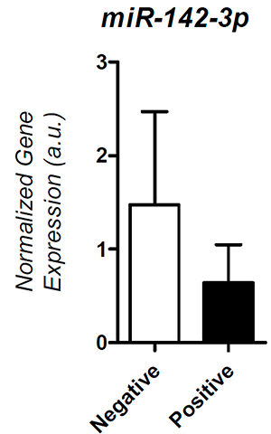
Figure 1. Expression of miR-142-3p in the Positive and Negative Implantation groups.
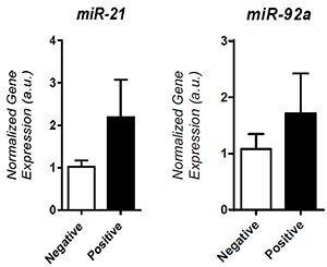
Figure 2. Expression of miR-21 and miR-92a in the Positive and Negative Implantation groups
DISCUSSION
The importance of miRNAs in embryonic stem cell lines and embryo development in several species has been proven. Several studies have described dynamic changes in miRNA expression in gametes and during early embryo development of mammalian species (Tang et al., 2007; Tesfaye et al., 2009; Hossain et al., 2012; Mondou et al., 2012; Abd El Naby et al., 2013). However, little is known about human embryo miRNA expression. The results from this pilot study demonstrate that increased expression of miR-142-3p might be associated with implantation failure in ICSI cycles.
Bioinformatic analysis of miR-142-3p predicted targets showed its potential role on the regulation of several biological pathways and cell functions, including cell cycle inhibition and tumor suppression (Lee et al., 2014). However, the biological role of miR-142-3p during early human embryo development is yet to be determined.
Hergenreider et al. (2012) showed that cultured cells secrete miRNA into the surrounding medium. In this context, embryos developing in vitro offer a promising possibility of detecting miRNA expression in spent culture medium and identifying noninvasive biomarkers of embryo viability and implantation potential.
The main characteristics of an ideal embryonic biomarker are: (1) noninvasive assessment, (ii) stability over time, (iii) embryo specificity, and (iv) easy measurement to allow fast assessment of embryo competence. MiRNAs seem to fit it perfectly (Capalbo et al., 2016). In fact, the stability of miRNAs and resistance to degradation is well known (Chen et al., 2008; Mraz et al., 2009). Since miRNAs may be encapsulated in exosomes, which are small vesicles that offer additional protection from degrading enzymes (Boon & Vickers, 2013) or conjugated with macromolecular complexes (Weber et al., 2010), they are protected from degradation and can be detected after extended periods of time (Jung et al., 2010).
Rosenbluth et al. (2013) were the first to attempt to describe miRNA secreted from embryos in culture medium. The authors observed that the most highly expressed miRNA in euploid embryos was miR-372. Several differentially expressed miRNAs were discovered based on chromosomal status, including gender of the embryo.
Another study by Rosenbluth et al, (2014) detected ten miRNAs, but eight yielded false-positive signals derived from the protein supplement used in the culture medium, since miRNAs were also present in the ‘blank’ culture medium prior to embryo culture. The authors also looked into whether MiRNAs were differentially secreted according to embryo chromosomal status and pregnancy outcome. Higher expression levels of two miRNAs, which were not present in the blank culture medium, were associated with embryo aneuploidy (miR-191) and pregnancy failure (miR-191 and miR-372). The association between both miRNAs and pregnancy failure was found only in embryos derived from conventional IVF cycles, suggesting that sperm injection alters miRNA secretion patterns.
More recently, Capalbo et al. (2016) comprehensively characterized the profile of miRNAs secreted by human embryos in spent culture medium and explored whether miRNAs could be used as biomarkers of ICSI outcomes. The study revealed that two miRNAs (miR-20a and miR-30c) had higher concentration levels in the spent medium of implanted blastocysts. Both miRNAs are suggested to be involved in 23 pathways related to embryo implantation.
Several different miRNAs have been involved in the assessment of embryo competence. However, there is no consensus in the literature over this issue. The inconsistencies may be explained by the different methods used in miRNA analysis and the differences in the day of collection of spent medium; in our study spent medium was collected on day 3 from embryos achieving the blastocyst stage on day 5, while others collected spent medium on day 5. Moreover, during embryo development, there is constant synthesis and degradation of miRNAs. It has been shown that miRNAs are maternally inherited with the loss of approximately 60% between the one- and two-cell stages during the maternal zygotic transition (Tang et al., 2007), with an overall increase in miRNAs expression by the blastocyst stage (Yang et al., 2008).
miRNAs have been suggested as suitable candidates for biomarkers of embryo competence for their association with several diseases (Bernstein et al., 2003; Tzur et al., 2008; Foshay & Gallicano, 2009; Medeiros et al., 2011; Wang et al., 2012). Recent studies indicated a biological role for miRNA in controlling ovarian function (Imbar and Eisenberg, 2014), in which there is intense exchange of miRNAs between the oocyte and granulosa cells (Fiedler et al., 2008). Some studies have also described altered miRNA expression in patients with ovarian dysfunctions, such as polycystic ovarian syndrome (Sang et al., 2013; Roth et al., 2014) and premature ovarian failure (Yang et al., 2012). Moreover, poor response to controlled ovarian stimulation has also been related to altered miRNA expression (Karakaya et al., 2015).
In animal models, new communication systems mediating the crosstalk between the preimplantation embryo and the endometrium based on miRNA expression have been recently discovered (Sengupta et al., 2006; Pawar et al., 2013; Rosario et al., 2014; Chu et al., 2015). Specific miRNAs may act by transferring information from the blastocyst to the surrounding endometrial cells, thus altering the outcome of implantation.
This study has been affected by the following limitations: (i) a small case basis, and (ii) a limited number of analyzed miRNAs. Despite the relatively small number of embryos included in our analysis, we were able to detect significant differences in miRNA expression between the groups. We confirmed that miR-142-3p is differentially expressed in human embryos that achieve the blastocyst stage according to their implantation status. This study provided additional evidence that miRNAs are secreted from human embryos into the culture medium, which makes miRNA a good candidate biomarker for embryo competence and implantation development. Future studies are required to determine whether embryo culture medium might be enriched or deprived of specific miRNAs and improve embryo development.
In conclusion, our preliminary results support the need to further explore miRNA expression in spent culture medium as a noninvasive biomarker of embryo quality and implantation potential in ICSI cycles.
REFERENCES
Abd El Naby WS, Hagos TH, Hossain MM, Salilew-Wondim D, Gad AY, Rings F, Cinar MU, Tholen E, Looft C, Schellander K, Hoelker M, Tesfaye D.. Expression analysis of regulatory microRNAs in bovine cumulus oocyte complex and preimplantation embryos. Zygote. 2013;21:31-51.
Medline Crossref
ACOG. ACOG Committee Opinion #324: Perinatal risks associated with assisted reproductive technology. Obstet Gynecol. 2005; 106: 1143-6.
Medline
Asirvatham AJ, Magner WJ, Tomasi TB. miRNA regulation of cytokine genes. Cytokine. 2009;45:58-69.
Medline Crossref
Barbarotto E, Schmittgen TD, Calin GA. MicroRNAs and cancer: profile, profile, profile. Int J Cancer. 2008; 122: 969-77.
Medline Crossref
Bernstein E, Kim SY, Carmell MA, Murchison EP, Alcorn H, Li MZ, Mills AA, Elledge SJ, Anderson KV, Hannon GJ. Dicer is essential for mouse development. Nat Genet. 2003;35:215-7.
Medline Crossref
Boon RA, Vickers KC. Intercellular transport of microRNAs. Arterioscler Thromb Vasc Biol. 2013;33:186-92.
Medline Crossref
Capalbo A, Ubaldi FM, Cimadomo D, Noli L, Khalaf Y, Farcomeni A, Ilic D, Rienzi L. MicroRNAs in spent blastocyst culture medium are derived from trophectoderm cells and can be explored for human embryo reproductive competence assessment. Fertil Steril. 2016;105:225-35.e1-3.
Medline Crossref
Carleton M, Cleary MA, Linsley PS. MicroRNAs and cell cycle regulation. Cell Cycle. 2007;6:2127-32.
Medline Crossref
Chen X, Ba Y, Ma L, Cai X, Yin Y, Wang K, Guo J, Zhang Y, Chen J, Guo X, Li Q, Li X, Wang W, Zhang Y, Wang J, Jiang X, Xiang Y, Xu C, Zheng P, Zhang J, Li R, Zhang H, Shang X, Gong T, Ning G, Wang J, Zen K, Zhang J, Zhang CY.Characterization of microRNAs in serum: a novel class of biomarkers for diagnosis of cancer and other diseases. Cell Res. 2008;18:997-1006.
Medline Crossref
Chu B, Zhong L, Dou S, Wang J, Li J, Wang M, Shi Q, Mei Y, Wu M. miRNA-181 regulates embryo implantation in mice through targeting leukemia inhibitory factor. J Mol Cell Biol. 2015;7:12-22.
Medline Crossref
Cortezzi SS, Cabral EC, Trevisan MG, Ferreira CR, Setti AS, Braga DP, Figueira Rde C, Iaconelli A Jr, Eberlin MN, Borges E Jr. Prediction of embryo implantation potential by mass spectrometry fingerprinting of the culture medium. Reproduction. 2013;145:453-62.
Medline Crossref
Cortezzi SS, Garcia JS, Ferreira CR, Braga DP, Figueira RC, Iaconelli A Jr, Souza GH, Borges E Jr, Eberlin MN. Secretome of the preimplantation human embryo by bottom-up label-free proteomics. Anal Bioanal Chem. 2011;401:1331-9.
Medline Crossref
de Mouzon J, Goossens V, Bhattacharya S, Castilla JA, Ferraretti AP, Korsak V, Kupka M, Nygren KG, Andersen AN; European IVF-Monitoring (EIM); Consortium for the European Society on Human Reproduction and Embryology (ESHRE). Assisted reproductive technology in Europe, 2007: results generated from European registers by ESHRE. Hum Reprod. 2012;27:954-66.
Medline Crossref
Fiedler SD, Carletti MZ, Hong X, Christenson LK. Hormonal regulation of MicroRNA expression in periovulatory mouse mural granulosa cells. Biol Reprod. 2008;79:1030-7.
Medline Crossref
Foshay KM, Gallicano GI. miR-17 family miRNAs are expressed during early mammalian development and regulate stem cell differentiation. Dev Biol. 2009; 326:431-43.
Medline Crossref
Hardarson T, Ahlström A, Rogberg L, Botros L, Hillensjö T, Westlander G, Sakkas D, Wikland M.Non-invasive metabolomic profiling of Day 2 and 5 embryo culture medium: a prospective randomized trial. Hum Reprod. 2012;27:89-96.
Medline Crossref
Hergenreider E, Heydt S, Tréguer K, Boettger T, Horrevoets AJ, Zeiher AM, Scheffer MP, Frangakis AS, Yin X, Mayr M, Braun T, Urbich C, Boon RA, Dimmeler S. Atheroprotective communication between endothelial cells and smooth muscle cells through miRNAs. Nat Cell Biol. 2012;14:249-56.
Medline Crossref
Hossain MM, Salilew-Wondim D, Schellander K, Tesfaye D. The role of microRNAs in mammalian oocytes and embryos. Anim Reprod Sci. 2012;134:36-44.
Medline Crossref
Imbar T, Eisenberg I. Regulatory role of microRNAs in ovarian function. Fertil Steril. 2014;101:1524-30.
Medline Crossref
Jung M, Schaefer A, Steiner I, Kempkensteffen C, Stephan C, Erbersdobler A, Jung K. Robust microRNA stability in degraded RNA preparations from human tissue and cell samples. Clin Chem. 2010;56:998-1006.
Medline Crossref
Karakaya C, Guzeloglu-Kayisli O, Uyar A, Kallen AN, Babayev E, Bozkurt N, Unsal E, Karabacak O, Seli E. Poor ovarian response in women undergoing in vitro fertilization is associated with altered microRNA expression in cumulus cells. Fertil Steril. 2015;103:1469-76.e1-3.
Medline Crossref
Lakshmipathy U, Love B, Goff LA, Jörnsten R, Graichen R, Hart RP, Chesnut JD. MicroRNA expression pattern of undifferentiated and differentiated human embryonic stem cells. Stem Cells Dev. 2007;16:1003-16.
Medline Crossref
Laurent LC. MicroRNAs in embryonic stem cells and early embryonic development. J Cell Mol Med. 2008;12:2181-8.
Medline Crossref
Lee RC, Feinbaum RL, Ambros V. The C. elegans heterochronic gene lin-4 encodes small RNAs with antisense complementarity to lin-14. Cell.1993;75:843-54.
Medline Crossref
Lee YY, Yang YP, Huang MC, Wang ML, Yen SH, Huang PI, Chen YW, Chiou SH, Lan YT, Ma HI, Shih YH, Chen MT.MicroRNA142-3p promotes tumor-initiating and radioresistant properties in malignant pediatric brain tumors. Cell Transplant. 2014;23:669-90.
Medline Crossref
McCallie B, Schoolcraft WB, Katz-Jaffe MG. Aberration of blastocyst microRNA expression is associated with human infertility. Fertil Steril. 2010;93:2374-82.
Medline Crossref
Medeiros LA, Dennis LM, Gill ME, Houbaviy H, Markoulaki S, Fu D, White AC, Kirak O, Sharp PA, Page DC, Jaenisch R. Mir-290-295 deficiency in mice results in partially penetrant embryonic lethality and germ cell defects. Proc Natl Acad Sci U S A. 2011;108:14163-8.
Medline Crossref
Mondou E, Dufort I, Gohin M, Fournier E, Sirard MA. Analysis of microRNAs and their precursors in bovine early embryonic development. Mol Hum Reprod. 2012;18:425-34.
Medline Crossref
Mouillet JF, Ouyang Y, Coyne CB, Sadovsky Y.MicroRNAs in placental health and disease. Am J Obstet Gynecol. 2015;213:S163-72
Medline Crossref
Mraz M, Malinova K, Mayer J, Pospisilova S. MicroRNA isolation and stability in stored RNA samples. Biochem Biophys Res Commun. 2009;390:1-4.
Medline Crossref
Pandian Z, Bhattacharya S, Ozturk O, Serour G, Templeton A.Number of embryos for transfer following in-vitro fertilisation or intra-cytoplasmic sperm injection. Cochrane Database Syst Rev. 2009; 2:CD003416.
Medline Crossref
Pawar S, Starosvetsky E, Orvis GD, Behringer RR, Bagchi IC, Bagchi MK. STAT3 regulates uterine epithelial remodeling and epithelial-stromal crosstalk during implantation. Mol Endocrinol. 2013;27:1996-2012.
Medline Crossref
Rosario GX, Hondo E, Jeong JW, Mutalif R, Ye X, Yee LX, Stewart CL.The LIF-mediumted molecular signature regulating murine embryo implantation. Biol Reprod. 2014;91:66.
Medline Crossref
Rosenbluth EM, Shelton DN, Sparks AE, Devor E, Christenson L, Van Voorhis BJ.Voorhis (2013). MicroRNA expression in the human blastocyst. Fertil Steril. 2013;99:855-861.e3.
Medline Crossref
Rosenbluth EM, Shelton DN, Wells LM, Sparks AE, Van Voorhis BJ. Human embryos secrete microRNAs into culture medium--a potential biomarker for implantation. Fertil Steril. 2014;101:1493-500.
Medline Crossref
Roth LW, McCallie B, Alvero R, Schoolcraft WB, Minjarez D, Katz-Jaffe MG. Altered microRNA and gene expression in the follicular fluid of women with polycystic ovary syndrome. J Assist Reprod Genet. 2014;31:355-62.
Medline Crossref
Sang Q, Yao Z, Wang H, Feng R, Wang H, Zhao X, Xing Q, Jin L, He L, Wu L, Wang L. Identification of microRNAs in human follicular fluid: characterization of microRNAs that govern steroidogenesis in vitro and are associated with polycystic ovary syndrome in vivo. J Clin Endocrinol Metab. 2013;98:3068-79.
Medline Crossref
Sengupta J, Lalitkumar PG, Najwa AR, Ghosh D. Monoclonal antileukemia inhibitory factor antibody inhibits blastocyst implantation in the rhesus monkey. Contraception. 2006;74:419-25.
Medline Crossref
Setti PE, Bulletti C. Strategies to improve embryo implantation to supraphysiological rates. Ann N Y Acad Sci. 2011;1221:75-9.
Medline Crossref
Suh N, Blelloch R. Small RNAs in early mammalian development: from gametes to gastrulation. Development. 2011;138:1653-61.
Medline Crossref
Sullivan CS, Ganem D. MicroRNAs and viral infection. Mol Cell. 2005;20:3-7.
Medline Crossref
Tang F, Kaneda M, O’Carroll D, Hajkova P, Barton SC, Sun YA, Lee C, Tarakhovsky A, Lao K, Surani MA.Maternal microRNAs are essential for mouse zygotic development. Genes Dev. 2007;21:644-8.
Medline Crossref
Tatsuguchi M, Seok HY, Callis TE, Thomson JM, Chen JF, Newman M, Rojas M, Hammond SM, Wang DZ.Expression of microRNAs is dynamically regulated during cardiomyocyte hypertrophy. J Mol Cell Cardiol. 2007;42:1137-41.
Medline Crossref
Tesfaye D1, Worku D, Rings F, Phatsara C, Tholen E, Schellander K, Hoelker M. Identification and expression profiling of microRNAs during bovine oocyte maturation using heterologous approach. Mol Reprod Dev. 2009;76:665-77
Medline Crossref
Thouas GA, Dominguez F, Green MP, Vilella F, Simon C, Gardner DK.Soluble ligands and their receptors in human embryo development and implantation. Endocr Rev. 2015; 36:92-130.
Medline Crossref
Tzur G, Levy A, Meiri E, Barad O, Spector Y, Bentwich Z, Mizrahi L, Katzenellenbogen M, Ben-Shushan E, Reubinoff BE, Galun E. MicroRNA expression patterns and function in endodermal differentiation of human embryonic stem cells. PLoS One. 2008;3:e3726.
Medline Crossref
Vergouw CG, Kieslinger DC, Kostelijk EH, Botros LL, Schats R, Hompes PG, Sakkas D, Lambalk CB. Day 3 embryo selection by metabolomic profiling of culture medium with near-infrared spectroscopy as an adjunct to morphology: a randomized controlled trial. Hum Reprod. 2012;27:2304-11.
Medline Crossref
Wang GK, Zhu JQ, Zhang JT, Li Q, Li Y, He J, Qin YW, Jing Q. Circulating microRNA: a novel potential biomarker for early diagnosis of acute myocardial infarction in humans. Eur Heart J. 2010;31:659-66.
Medline Crossref
Wang K, Zhang S, Weber J, Baxter D, Galas DJ. Export of microRNAs and microRNA-protective protein by mammalian cells. Nucleic Acids Res. 2010b;38:7248-59.
Medline Crossref
Wang R, Hu Y, Song G, Hao CJ, Cui Y, Xia HF, Ma X. MiR-206 regulates neural cells proliferation and apoptosis via Otx2. Cell Physiol Biochem. 2012;29:381-90.
Medline Crossref
Weber JA, Baxter DH, Zhang S, Huang DY, Huang KH, Lee MJ, Galas DJ, Wang K. The microRNA spectrum in 12 body fluids. Clin Chem. 2010;56:1733-41.
Medline Crossref
Wightman B, Ha I, Ruvkun G. Posttranscriptional regulation of the heterochronic gene lin-14 by lin-4 mediumtes temporal pattern formation in C. elegans. Cell. 1993; 75:855-62.
Medline Crossref
Yang X, Zhou Y, Peng S, Wu L, Lin HY, Wang S, Wang H.Differentially expressed plasma microRNAs in premature ovarian failure patients and the potential regulatory function of mir-23a in granulosa cell apoptosis. Reproduction. 2012;144:235-44.
Medline Crossref
Yang Y, Bai W, Zhang L, Yin G, Wang X, Wang J, Zhao H, Han Y, Yao YQ. Determination of microRNAs in mouse preimplantation embryos by microarray. Dev Dyn. 2008;237:2315-2.
Medline Crossref