JBRA Assist. Reprod. 2016;20 (4):212-216
ORIGINAL ARTICLE
doi: 10.5935/1518-0557.20160041
The impact of male age on embryo quality: a retrospective study using time-lapse imaging
1Embryolife Reproductive Medicine Institute, São José dos Campos/SP
CONFLICT OF INTERESTS
The author declares that she has no conflict of interest.
ABSTRACT
Objective: This study aimed to correlate male age with embryo morphokinetic parameters on D3 considering the timing and the exact moment of embryo cleavage.
Methods: Time-lapse imaging was used to produce an ideal cleavage curve for the embryos analyzed. The percentage of embryos under the curve was analyzed and correlated with male age.
Results: 32.6% of the embryos from patients aged 28-33 years were under the curve; 36.2% of the embryos from patients aged 34-39 years were under the curve; 41.3% of the embryos from patients aged 40-45 years were under the curve; and 26.3% of the embryos fro patients aged 46-57 years were under the curve.
Conclusions: a statistically non-significant decrease was observed in the percentage of embryos under the optimal cleavage curve on D3 in the group of men aged between 40 and 45 years. Further studies looking into embryos in the blastocyst stage (D5 or D6) are required.
Keywords: Male age, time-lapse, morphokinetics, embryo quality.
INTRODUCTION
Male age has been associated with decreased semen quality (Schwartz et al., 1983; Bujan et al., 1988; Silva et al., 2012; Oliveira et al., 2014.). It is also known that advanced paternal age is linked to higher rates of miscarriage (de la Rochebrochard & Thonneau., 2002; Slama et al., 2005; Kleinhaus et al., 2006), autosomal dominant diseases, aneuploidy, and other diseases (Glaser et al., 2003; Schmid et al., 2007). Other authors have associated advanced male age with greater sperm DNA fragmentation (Simon et al., 2014).
Abnormal sperm morphology and changes in embryo morphology have been closely linked, suggesting that sperm quality affects embryo development (Vagnini et al., 2007; Meng et al., 2016.).
Time-lapse imaging has been used to monitor embryo development and help pick the best embryo based on cleavage kinetics (Meseguer et al., 2011; Cruz et al., 2012.; Herrero et al., 2013; Kirkegaard et al., 2012; 2013; Aguilar et al., 2014; Basile et al., 2015). This imaging technique accurately predicts blastocyst formation (Motato et al., 2016), allowing early selection of embryos with high implantation potential within shorter periods of incubation (Milewski et al., 2015). Interestingly, prolonged embryo culture has been associated with significant epigenetic changes (Lonergan et al., 2003; Calle et al., 2012) and increased risk of preterm delivery when compared to embryos transferred on D2 or D3 (Maheshwari et al., 2013; Giving et al., 2014).
Considering the timing and exact moment of embryo cleavage described by Meseguer et al., 2011, this study aimed to find whether male age correlated with embryo morphokinetic parameters on D3.
MATERIALS AND METHODS
Two hundred and ninety-six embryos obtained from intracytoplasmic sperm injection (ICSI) procedures were included in the study.
The embryos were analyzed using time-lapse imaging (10/10 min), and the exact time of occurrence of significant embryo development events was noted.
ICSI
ICSI was performed on culture medium containing HEPES. A Nikon® Eclipse TE 2000-S microscope at 250x magnification was used. Temperature was controlled in the central vinyl surface of the micro-handler table with a Greisinger® GMH 3230 surface thermometer (Germany) with validated calibration. After ICSI, the embryos were rinsed with the same culture medium in which they developed. Rinsing was carried out with at least three drops (~ 50mL) of pre-equilibrated medium. Then the oocytes were placed in micro-wells from the special time-lapse board and taken to an incubator.
Incubation
The same culture medium was used for all embryos included in this study (Basile -., 2013). The CO2 level was as indicated by the manufacturer of the medium, while O2 levels were kept at ~ 20%.
Culture plates with nine or sixteen wells were prepared and pre-equilibrated in the incubator. After pre-equilibration, all micro-bubbles were carefully removed.
Image acquisition system
The images were captured with a microscope camera placed inside a “big box” incubator type. Photos were taken every 10 minutes for the composition of a time line. The system used a green homogeneous LED light source.
Morphokinetic parameter assessment based on time-lapse imaging
A software program was used to retrospectively analyze the images depicting the events that occurred after ICSI, and identify the precise moments at which pronuclei and cell walls disappeared and abnormalities arose.
An ideal cleavage curve plotted with the aid of analysis software was considered (Meseguer et al., 2011).
Graphic 1 illustrates embryo cleavage. A slight delay was observed in cleavage from three to four cells, which was enough to distinguish the embryos falling outside the optimal development curve.
Female factor infertility
In order to mitigate the impact of female factor infertility, only the data from oocytes not presenting morphological abnormalities were analyzed (REDLARA 2006).
Statistical analysis
Quantitative variables were described by means of measures of central tendency, scatter, and position, whereas male age was categorically described in terms of absolute and relative frequencies.
Binomial logistic regression with robust variance was used to check the impact of male age on the number of embryos (Mccullagh & Nelder, 1989). The software used in the analysis was the R (version 3.2.2).
RESULTS
Table 1 shows the description of the studied variables.
• Mean age was 37.88 years, with a standard deviation of 6.87 years.
• A mean of 1.79 embryos were under the curve; the minimum and maximum values were 0 and 9, respectively.
• The number of embryos ranged from 1 to 15; the mean number of embryos was 5.10.
• A mean of 35.1% of the embryos were under the curve.
Table 2 shows the percentage of embryos under the age curve; in it, 41.3% of the embryos of patients aged between 40 and 45 years were under the curve, versus 26.3% of the embryos of patients aged between 46 and 57 years.
Table 3 shows the binomial logistic regression with robust variance (Mccullagh & Nelder, 1989) adjusted to check for the impact of male age on the number of embryos under the curve. The following conclusions may be derived:
• The chance of an individual aged 34-39 having an embryo under of the curve was 1.17 [0.53; 2.59] times the chance of an individual aged 28-33 years, but this difference was not statistically significant (P-value = 0.696).
• The chance of an individual aged 40-45 having an embryo under of the curve was 1.46 [0.64; 3.34] times the chance of an individual aged 28-33 years, but this difference was not statistically significant (P-value = 0.377).
• The chance of an individual aged 46-57 having an embryo inside of the curve was 0.76 [0.25; 2.22] times the chance of an individual aged 28-33 years, but this difference was not statistically significant (P-value = 0.592).
A comparison against the findings on Table 3 shows the following:
• The chance of an individual aged 40-45 having an embryo under of the curve was 1.24 [0.54; 2.84] times the chance of an individual aged 34-39 years, but this difference was not statistically significant (P-value = 0.600).
• The chance of an individual aged 46-57 having an embryo inside of the curve was 0.63 [0.21; 1.91] times the chance of an individual aged 34-39 years, but this difference was not statistically significant (P-value = 0.408).
• The chance of an individual aged 46-57 having an embryo inside of the curve was 0.51 [0.16; 1.58] times the chance of an individual aged 40-45 years, but this difference was not statistically significant (P-value = 0.236).
Graph 2 shows the percentage of embryos under the curve for each age group with P-values estimated by binomial logistic regression with robust variance, as shown in Table 3.
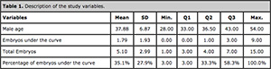
Table 1. Description of the study variables.
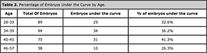
Table 2. Percentage of Embryos Under the Curve by Age.
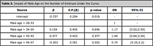
Table 3. Impact of Male Age on the Number of Embryos Under the Curve.
DISCUSSION
Various different aspects concerned with the impact of male age on semen quality have been described in the literature. Some studies have shown an inverse correlation between male age and semen volume – volume decreasing with age – (Spandorfer et al., 1998; Andolz et al., 1999; Moskovtsev et al., 2009; Brahem et al., 2011; Oliveira et al., 2014), sperm motility (Moskovtsev et al., 2009; Brahem et al., 2011; Dain et al., 2011; Stone et al., 2013; Oliveira et al., 2014) and sperm vitality (Moskovtsev et al., 2009; Brahem et al., 2011; Zhu et al., 2011; Stone et al., 2013). Conversely, other authors failed to observe connections between any such semen parameters and paternal age (Berling et al., 1997; Spandorfer et al., 1998; Frattarelli et al., 2008; Nijs et al., 2011; Fréour et al., 2012). Some studies found no correlation between male age and semen concentration (Spandorfer et al., 1998; Frattarelli et al., 2008; Bellver et al., 2008; Dain et al., 2011; Nijs et al., 2011; Fréour et al., 2012), whereas other authors have either described decreases (Luna et al., 2009; Stone et al., 2013) or increases (Andol et al., 1999; Brahem et al., 2011) in semen concentration over time. The studies cited above generally differ over their conclusions and some do not specify a number of points such as whether the group of enrolled patients includes solely individuals seen at ART clinics, how many of them smoke or drink alcohol, have varicocele, or are taking medication or vitamins. These differences complicate the interpretation of results.
Discrepant findings have been reported in the literature in regards to sperm nuclear vacuoles. Some studies have identified significant correlations between male age and sperm nuclear vacuoles (Braga et al., 2011; Silva et al., 2012; Oliveira et al., 2014). However, two of the authors (Braga et al., 2011; Silva et al., 2012) reported that there was no correlation between normal sperm frequency and male age defined by MSOME (motile sperm organelle morphology examination).
Authors correlating paternal age and embryo development have claimed that embryo morphology during cleavage is not affected by male age (Frattarelli et al., 2008) and that male age is irrelevant for the outcome ART procedures (Bellver et al., 2008), while others believe there is not enough data to support such claim (Dain et al., 2011). However, a significant decrease in blastocyst formation was observed with increasing age (Luna et al., 2009; Dain et al., 2011), probably reflecting the paternal genome activation in the embryo.
All previous articles analyzed male age in relation to embryo quality by considering exclusively embryo morphology criteria. It is important to remember that morphologically identical embryos may be assessed or fall into the exclusion criteria according to the algorithm proposed by Basile et al. (2015). Events related to low implantation rates such as multinucleation (Pickering et al., 1995), asymmetric blastomeres (Hardarson et al., 2001), direct cleavage to three cells (1C-3C) (Rubio et al., 2012) and asynchronous disappearance of pronuclei (Rosário et al., 2015) may be difficult or impossible to observe without the aid of time-lapse imaging.
Table 2 and Graph 2 in this study describe decreased percentages of embryos under the normal cleavage curve, as also shown by other authors. This finding was noted in patients approaching the fifth decade of life. However, statistical tests showed that such decrease was not significant. This finding was also reported in other studies (Gallardo et al., 1996; Aboulghar et al., 2007; Frattarelli et al., 2008; Bellver et al., 2008; Dain et al., 2011).
Semen quality may decrease with advanced age, but the actual impact of male age on embryo viability is multifactorial.
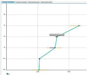
Graph 1. Embryo development curve.
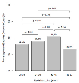
Graph 2. Percentage of embryos under the curve for each age group
CONCLUSION
Decreased percentages of embryos under the normal cleavage curve on D3 were found for males aged 45 years and older, but such difference was not statistically significant. Further studies are required to assess the status of embryos on the blastocyst stage (D5 or D6).
REFERENCES
Aboulghar M, Mansour R, Al-Inany H, AbouSetta AM, Aboulghar M, Mourad L. Paternal age and outcome of intracytoplasmic sperm injection. Reprod Biomed Online 2007;14:588–92.
Medline Crossref
Aguilar J, Motato Y, Escriba MJ, Ojeda M, Munoz E, Meseguer M. The human first cell cycle: impact on implantation. Reprod Biomed Online 2014; 28:475– 84.
Medline Crossref
Andolz P, Bielsa MA, Vila J. Evolution of semen quality in North-eastern Spain: a study in 22,759 infertile men over a 36 year period. Hum Reprod 1999;14:731–35.
Medline Crossref
Basile N, Morbeck D, García-Velasco J, Bronet F, Meseguer M.Type of culture media does not affect embryo kinetics: a time-lapse analysis of sibling oocytes. Hum Reprod. 2013;28:634-41.
Medline Crossref
Basile N, Vime P, Florensa M, Aparicio Ruiz B, García Velasco JA, Remohí J, Meseguer M.The use of morphokinetics as a predictor of implantation: a multicentric study to define and validate an algorithm for embryo selection. Hum Reprod. 2015;30:276-83.
Medline Crossref
Bellver J, Garrido N, Remohí J, Pellicer A, Meseguer M. Influence of paternal age on assisted reproduction outcome. Reprod Biomed Online 2008;17:595– 604.
Medline Crossref
Berling S, Wölner-Hanssen P. No evidence of deteriorating semen quality among men in infertile relationships during the last decade: a study of males from Southern Sweden. Hum Reprod 1997;12:1002–5.
Medline Crossref
Braga DPAF, Setti AS, Figueira RCS, Nichi M, Martinhago CD, Iaconelli A, Borges E. Sperm organelle morphologic abnormalities: Contributing factors and effects on intracytoplasmic sperm injection cycles outcomes. Urology 2011;78:786–91.
Medline Crossref
Brahem S, Mehdi M, Elghezal H, Saad A. The effects of male aging on semen quality, sperm DNA fragmentation and chromosomal abnormalities in an infertile population. J Assist Reprod Genet 2011;28:425–32.
Medline Crossref
Bujan L, Mieusset R, Mondinat C, Mansat A, Pontonnier F: Sperm morphology in fertile men and its age related variation. Andrologia 1988, 20:121-8.
Medline Crossref
Calle A, Fernandez-Gonzalez R, Ramos-Ibeas P, Laguna-Barraza R, Perez- Cerezales S, Bermejo-Alvarez P, Ramirez MA, Gutierrez-Adan A. Long-term and transgenerational effects of in vitro culture on mouse embryos. Theriogenology. 2012:77:785-93.
Medline Crossref
Cruz M, Garrido N, Herrero J, Perez-Cano I, Munoz M, Meseguer M. Timing of cell division in human cleavage-stage embryos is linked with blastocyst formation and quality. Reprod Biomed Online 2012;25:371–81.
Medline Crossref
Dain L, Auslander R, Dirnfeld M. The effect of paternal age on assisted reproduction outcome. Fertil Steril 2011;95:1–8.
Medline Crossref
de la Rochebrochard E, Thonneau P. Paternal age and maternal age are risk factors for miscarriage; results of a multicentre European study. Hum Reprod 2002, 17:1649-56.
Medline Crossref
Frattarelli JL, Miller KA, Miller BT, Elkind-Hirsch K, Scott RT. Male age negatively impacts embryo development and reproductive outcome in donor oocyte assisted reproductive technology cycles. Fertil Steril 2008;90:97–103.
Medline Crossref
Fréour T, Jean M, Mirallie S, Barriere P. Computer-assisted sperm analysis parameters in young fertile sperm donors and relationship with age. Syst Biol Reprod Med. 2012;58:102–6.
Medline Crossref
Gallardo E, Simón C, Levy M, Guanes PP, Remohí J, Pellicer A. Effect of age on sperm fertility potential: oocyte donation as a model. Fertil Steril. 1996;66:260–4.
Medline
Glaser RL, Broman KW, Schulman RL, Eskenazi B, Wyrobek AJ, Jabs EW: The paternal-age effect in Apert syndrome is due, in part, to the increased frequency of mutations in sperm. Am J Hum Genet. 2003;73:939-47.
Medline Crossref
Hardarson T, Hanson C, Sjögren A, Lundin K. Human embryos with unevenly sized blastomeres have lower pregnancy and implantation rates: indications for aneuploidy and multinucleation. Hum Reprod. 2001;16:313–8.
Medline Crossref
Herrero J, Tejera A, Albert C, Vidal C, de los Santos MJ, Meseguer M. A time to look back: analysis of morphokinetic characteristics of human embryo development. Fertil Steril. 2013;100:1602-9.e1-4.
Medline Crossref
Kirkegaard K, Agerholm IE, Ingerslev HJ. Time-lapse monitoring as a tool for clinical embryo assessment. Hum Reprod. 2012;27:1277-85.
Medline Crossref
Kirkegaard K, Kesmodel US, Hindkjaer JJ, Ingerslev HJ. Time-lapse parameters aspredictorsof blastocyst development and pregnancy outcome in embryos from good prognosis patients: a prospective cohort study. Hum Reprod. 2013; 28:2643–51.
Medline Crossref
Kleinhaus K, Perrin M, Friedlander Y, Paltiel O, Malaspina D, Harlap S: Paternal age and spontaneous abortion. Obstet Gynecol. 2006, 108:369-77.
Medline Crossref
Lonergan P, Rizos D, Gutierrez-Adan A, Fair T, Boland MP. Effect of culture environment on embryo quality and gene expression-experience from animal studies. Reprod Biomed Online. 2003;7:657-63.
Medline Crossref
Luna M, Finkler E, Barritt J, Bar-Chama N, Sandler B, Copperman AB, Grunfeld L. Paternal age and assisted reproductive technology outcome in ovum recipients. Fertil Steril. 2009;92:1772–75.
Medline Crossref
Maheshwari A, Kalampokas T, Davidson J, Bhattacharya S. Obstetric and perinatal outcomes in singleton pregnancies resulting from the transfer of blastocyst-stage versus cleavage-stage embryos generated through in vitro fertilization treatment: a systematic review and meta-analysis. Fertil Steril. 2013;100:1615-21.e1-10.
Medline Crossref
Mccullagh P, Nelder JA, eds. Generalized Linear Models. Chapman and Hall/CRC press; 1989.
Meng XQ, Gong Y, Huang J, Zeng YM, Quan S, Zhong Y. Impact of sperm midpiece morphology on embryo development following intracytoplasmic morphologically selected sperm injection. Nan Fang Yi Ke Da Xue Xue Bao. 2016;36:255-9.
Medline
Meseguer M, Herrero J, Tejera A, Hilligsoe KM, Ramsing N, Remohi J. The use of morphokinetics as a predictor of embryo implantation. Hum Reprod. 2011;26:2658–71.
Medline Crossref
Milewski R, Kuć P, Kuczyńska A, Stankiewicz B, Łukaszuk K, Kuczyński W. A predictive model for blastocyst formation based on morphokinetic parameters in time-lapse monitoring of embryo development. J Assist Reprod Genet. 2015;32:571-9.
Medline Crossref
Moskovtsev SI, Willis J, White J, Mullen JBM. Sperm DNA Damage: Correlation to Severity of Semen Abnormalities. Urology 2009;74:789–93.
Medline Crossref
Motato Y, de los Santos MJ, Escriba MJ, Ruiz BA, Remohí J, Meseguer M. Morphokinetic analysis and embryonic prediction for blastocyst formation through an integrated time-lapse system. Fertil Steril 2016;105:376- 84.e9.
Medline Crossref
Nijs M, Jonge C De, Cox A, Janssen M, Bosmans E, Ombelet W. Correlation between male age, WHO sperm parameters, DNA fragmentation, chromatin packaging and outcome in assisted reproduction technology. Andrologia 2011; 43:174–9.
Medline Crossref
Oliveira J B A, Petersen C G, Mauri A L, Vagnini L D, Baruffi R L R, Franco Jr J G. The effects of age on sperm quality: an evaluation of 1,500 semen samples. JBRA Assist Reprod. 2014; 18:34-41.
Crossref
Pickering SJ, Taylor A, Johnson MH, Braude PR. An analysis of multinucleated blastomere formation in human embryos. Hum Reprod. 1995;10:1912–22.
Medline
RED LARA- Red Latinoamericana De Reproducción Asistid, ed. Manual de Procedimentos : Laboratório de Reprodução Assistida, 2006. p53-4.
Link
Rosário GR, Franco AC, Silva AV Asynchronous Detected Disappearance of the Pro-nuclei by Time-Lapse X Embryonic Development. JBRA Assist Reprod. 2015;19:114-8.
Medline Crossref
Rubio I, Kuhlmann R, Agerholm I, Kirk J, Herrero J, Escriba MJ, Bellver J, Meseguer M. Limited implantation success of direct-cleaved human zygotes: a time-lapse study. Fertil Steril.2012;98:1458–63.
Medline Crossref
Schmid TE, Eskenazi B, Baumgartner A, Marchetti F, Young S, Weldon R, Anderson D, Wyrobek AJ. The effects of male age on sperm DNA damage in healthy non-smokers. Hum Reprod. 2007; 22:180-7.
Medline Crossref
Schwartz D, Mayaux MJ, Spira A, Moscato ML, Jouannet P, Czyglik F, David G. Semen characteristics as a function of age in 833 fertile men. Fertil Steril. 1983; 39:530-5.
Medline
Silva LFI, Oliveira JB, Petersen CG, Mauri AL, Massaro FC, Cavagna M, Baruffi RLR, Franco JG. The effects of male age on sperm analysis by motile sperm organelle morphology examination (MSOME). Reprod Biol Endocrinol 2012;10:19.
Medline Crossref
Simon L, Murphy K, Shamsi MB, Liu L, Emery B, Aston KI, Hotaling J, Carrell DT. Paternal influence of sperm DNA integrity on early embryonic development. Hum Reprod. 2014;29:2402-12.
Medline Crossref
Slama R, Bouyer J, Windham G, Fenster L, Werwatz A, Swan SH: Influence of paternal age on the risk of spontaneous abortion. Am J Epidemiol. 2005; 161:816-23.
Medline Crossref
Spandorfer SD, Avrech OM, Colombero LT, Palermo GD, Rosenwaks Z. Effect of parental age on fertilization and pregnancy characteristics in couples treated by intracytoplasmic sperm injection. Hum Reprod. 1998;13:334–8.
Medline Crossref
Stone BA, Alex A, Werlin LB, Marrs RP. Age thresholds for changes in semen parameters in men. Fertil Steril 2013;100:952–8.
Medline Crossref
Vagnini L, Baruffi RL, Mauri AL, Petersen CG, Massaro FC, Pontes A, Oliveira JB, Franco JG Jr. The effects of male age on sperm DNA damage in an infertile population. Reprod Biomed Online. 2007;15:514-9.
Medline Crossref
Zhu QX, Meads C, Lu ML, Wu JQ, Zhou WJ, Gao ES.Turning point of age for semen quality: a population-based study in Chinese men. Fertil Steril 2011;96:572–6.
Medline Crossref