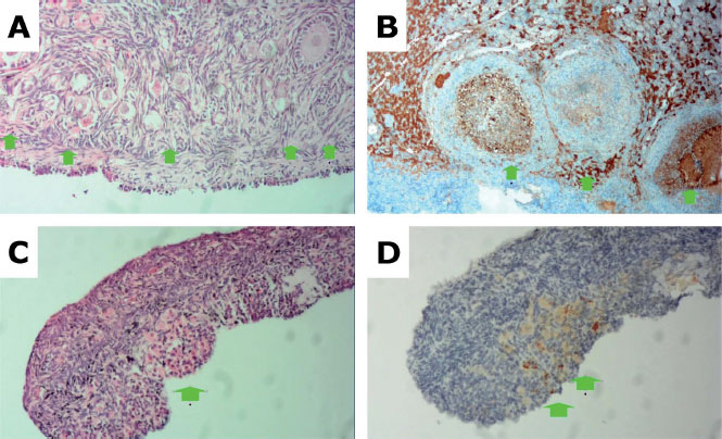
Figure 1. (A) Fresh ovarian germinal tissue specimen stained by HE (rabbit 5) showing multiple follicles. (B) Immunohistochemistry staining of fresh ovarian germinal tissue specimen showing several follicles (arrows) with strong Bcl-2 staining in sepia color (3+). (C) HE-stained germ cell solution (solution 3) revealing the presence of ovarian tissue (elongated stromal cells) and the outline of a follicle (arrow). (D) Immunohistochemistry staining of germ cell solution with weak sepia Bcl-2 staining (2+).