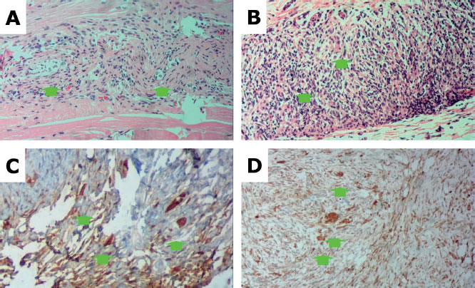
Figure 2. (A) Excisional biopsy specimen from a rabbit given a homologous graft (Rabbit 3) stained with HE showing ovarian tissue containing stroma and a few primordial follicles (arrows) among the soft tissue, absence of necrosis or inflammatory cells. (B) Excisional biopsy specimen from a rabbit given a heterologous graft (rabbit 4) stained with HE, showing ovarian tissue containing stroma and a few primordial follicles among the soft tissue, absence of necrosis, and presence of inflammatory cells. (C) Excisional biopsy specimen from a rabbit given a homologous graft (rabbit 3) with Bcl-2 staining, showing strong sepia marking (3+) in the cell cytoplasm. (D) Excisional biopsy specimen from a rabbit given a heterologous graft (rabbit 4) with Bcl-2 staining showing strong marking (3+) in sepia color in the cell cytoplasm.