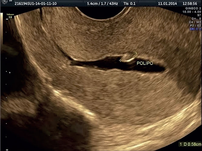
Figure 1. The distention of the uterine cavity produced after the infusion of saline solution allows for better visualization of pathologies. An endometrial polyp measuring 5.8 mm can be seen on the back wall.
Figure 1. The distention of the uterine cavity produced after the infusion of saline solution allows for better visualization of pathologies. An endometrial polyp measuring 5.8 mm can be seen on the back wall.