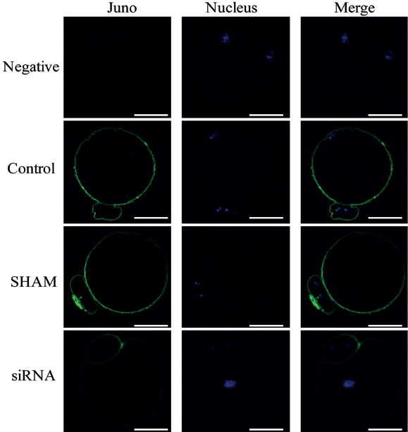
Figure 5. Immunofluorescence staining of MII stage oocyte after siRNA microinjection during GV stage. Juno was stained in green and nucleus was in blue. Scale bar=25 µm
Figure 5. Immunofluorescence staining of MII stage oocyte after siRNA microinjection during GV stage. Juno was stained in green and nucleus was in blue. Scale bar=25 µm