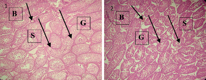
Figure 2. Plate 1- Photomicrograph showing the testes of control pups with normal seminiferous tubules and distinct peritubular tissue (S), with intact and adequate germ cells and spermatids (G). Blood vessels are also present (B). Plate 2- Photomicrograph showing the testes of nicotine-treated pups with apparently atrophic and indistinct seminiferous tubules (S). It also shows gradual erosion of the germ cell layer (G). Blood vessels are also present (B).