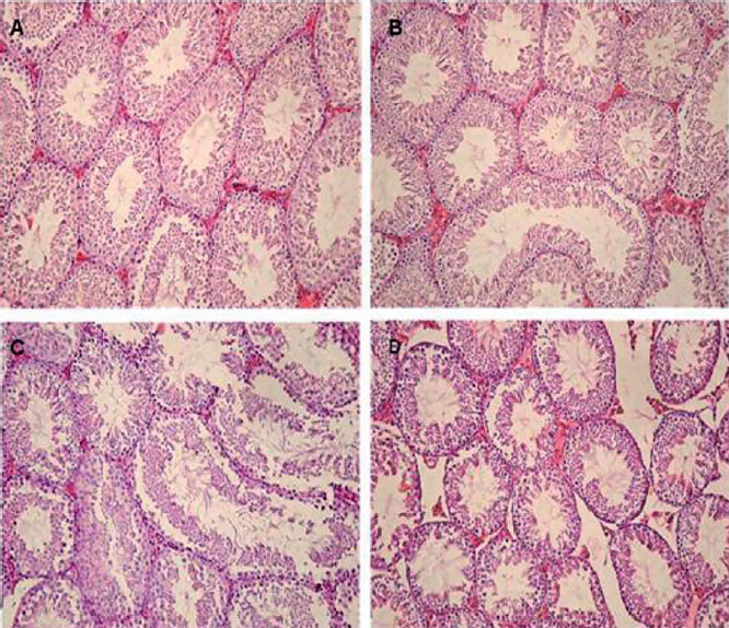
Figure 1. Light microscopy of cross sections of H & E stained testis from Control and Experimental groups. (A) Control testis: normal architecture of the seminiferous tubules is found. (B) 75mg/kg group, the tubules show normal spermatogenesis. (C) 375mg/kg group, there are histopathological changes including detachment, sloughing, and vacuolization. (D) Tubules show varying degrees of histopathological changes, and an increase in interstitial spaces between seminiferous tubules due to reduction in tubule diameter. (Magnifications: 40×).