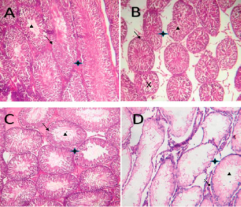
Plate 1. Photomicrographs of testicular tissue showing seminiferous tubules that are lined with germ cells (black arrow) at various stages of maturation to spermatozoa (black triangular box) and their respective interstitial spaces (black star box).
(A) Distilled water only (B) Cyclophosphamide only (C) N-acetylcysteine only (D) Cyclophosphamide + N-acetylcysteine.