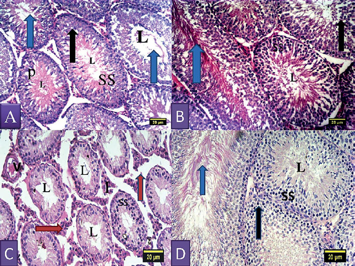
Figure 2. Section of the testis of control rat showing the seminiferous tubules containing cells of the spermatogenic series (SS) and the lumen (L) containing spermatozoa; Black arrow represents spermatogonium; P represents primary spermatocytes; Blue arrow represents spermatids and spermatozoa. (A-B) Section of the testis of rat treated with nitrite showing hypocellularity, reduction in cells of the spermatogenic series (SS) as a result of degeneration, sloughing and shortening of seminiferous epithelium; The seminiferous tubules show a single layer of basal spermatogonia; widened empty lumen (L); widened interstitium (I) due to tubular atrophy as a result of degeneration, Leydig cells show hyperplasia (brown arrows) and V shows vascular hemorrhage. (C) Section of the testis of rat treated with nitrite and walnut oil showing cells of the spermatogenic series and normal cellular composition in their germinal epithelium with sperm cells in the lumen and a normal interstitium. (H&E; X100).