JBRA Assist. Reprod. 2019;23(4):333-335
ORIGINAL ARTICLE SHORT COMUNICATION
doi: 10.5935/1518-0557.20190004
Endometriosis III and IV as a risk factor for tubal obstruction in infertile women
1Reproduction Human Laboratory. Obstetrics and Gynecology Dept. Federal University of Goiás State, Brazil
2Nutrition Academic, Federal University of Goias State, Brazil
CONFLICT OF INTEREST
The authors have no conflict of interest to declare.
ABSTRACT
Objective: A previous study carried out among infertile women with tubal obstruction identified a relative risk of 2.5 for Chlamydia trachomatis seropositivity. However, endometriosis may also be associated with increased risk. This study aimed to evaluate the risk of tubal obstruction associated with endometriosis III/IV among women submitted to assisted reproductive procedures.
Methods: A case-control study was performed among 144 women with and without tubal obstruction. We calculated the odds ratio with 95% CI regarding the association of endometriosis III/IV and tubal obstruction. Calculations were performed using the SPSS v.17.0 package.
Results: The mean age was 33.7 years (4.76 SD), and the mean infertility duration time was 66.7 months (120.6 SD). The total prevalence of endometriosis was 20/144 (13%). Among 144 women, the risk group with tubal obstruction and endometriosis III/IV comprised 7out of 20 (35%), compared with the group without such risk, that comprised 22 out of 124 (17%). The X2 test was 3.19 with a p-value of 0.07. The odds ratio (OR) was 2.5 (95% CI: 0.647-9.639).
Conclusion: Although the OR was 2.5, there was no significant difference between the groups with and without endometriosis III/IV. Further studies are needed to increase the sample size.
Keywords: endometriosis, tubal obstruction, tubal occlusion, infertility
INTRODUCTION
The prevalence of infertility in patients with endometriosis varies depending on the author. Some have reported that 25-50% of women with infertility have endometriosis, and about 30-50% of the women with endometriosis have infertility (Hickey et al., 2014). Between 10 and 15 % of all women seek IVF in the UK due to tubal infertility (HFEA, 2016). In this cases, the cause may be pelvic inflammatory disease, endometriosis, salpingitis isthmica nodosa, polyps and surgical trauma (Honoré et al., 1999). Tanahatoe et al. (2003) found 10 % of infertility in endometriosis. These authors also found that the most important causes of infertility in couples are ovulation disorders, tubal obstruction and semen abnormalities (mainly azoospermia, oligozoospermia, teratozoospermia and astenozoospermia). This causes account for approximately 75% of infertility in couples. The remaining is so far unknown (Tanahatoe et al., 2003). Mild or moderate endometriosis is related to subfertility with pregnancy rates of 17.7% at nine months of follow-up. Endometriosis is a net factor of subfertility, mainly in stages III and IV. In a series of cases, a fertility rate of 3% has been reported after 12 months in cases of stage IV endometriosis (Marcoux et al., 1997).
It is a disease that can affect several organs, such as the pelvic peritoneum, fallopian tubes, ovaries, subcutaneous tissue, umbilicus, urinary tract, bladder, heart, kidney, lung, liver, pancreas, muscles, central nervous system, among others, which makes it a multi-systemic disease (Goldberg & Bedaiwy, 2007; Lee et al., 2008). Endometriotic lesions are more frequent in the peritoneum and pelvic organs, especially in the ovaries, followed by the recto-vaginal septum. It is found less frequently in extra-pelvic regions, such as gastrointestinal (sigmoid, rectum, ileocecal and appendix) and urinary tract, extremities, subcutaneous tissue and abdominal wall (Lee et al., 2008). The mechanism of impaired fertility in endometriosis may involve anatomical distortions in the pelvis, adhesions, endometriomas or the production of substances (prostaglandins, cytokines, and growth factors) that are harmful to normal ovarian function, ovulation, fertilization and implantation. The really valid mechanisms are tubal obstruction, pelvic adhesions and ovarian endometriomas that distort anatomical relationships, limit the access of oocytes and spermatozoa and alter fimbriae mobility, mainly in stages III and IV (Mahutte & Arici, 2002). Phenomena such as anovulation, endocrine dysfunction, luteinized unruptured follicle syndrome, inadequate luteal phase, autoimmune dysfunction, abnormalities of the ovule quality and sperm alterations are theoretical mechanisms, still unproven, used to explain infertility in endometriosis in stages I and II (Toya et al., 2000). However, the two most probable mechanisms to explain the infertility in these stages are maturing on the late follicular phase and the antispermatic effect impairing folliculogenesis with oocyte alterations.
There are very few publications about the effect of endometriosis on tubal permeability. Some time ago Bowman & Cooke (1994) found that there was a strong correlation between the degree of intratubal damage and the extent of pelvic adhesions when the etiology was a previous pelvic inflammatory disease (PID), but not when the underlying etiology was endometriosis. However, in the endometriosis subgroup, intraluminal ampullary pathology was noted in 3 of 11 tubes (27%) assessed, and intraluminal fimbrial pathology was noted in 4 of 11 tubes (36%) assessed.
Osuga et al. (2008) describe a case of a patient with endometriosis who sought infertility treatment. During ovarian stimulation, an image of hydrosalpinx without infection appears and changed dramatically in size with the menstrual cycle. The patient was 32 years old and had had endometriosis since 24 years of age. She underwent ethanol sclerotherapy of a bilateral ovarian endometrioma at age 26 and laparoscopic cystectomy for ovarian endometrioma at age 30. Serum Chlamydia trachomatis IgA and IgM antibodies were negative. During ultrasonography work-up to check follicular growth and ovulation, the author noticed a hydrosalpinx-like structure that appeared larger at each ultrasound scan. This structure was minimal during the menstrual period. It would reach its maximum size during ovulation, and then shrank again. A later laparoscopy revealed endometriosis and tubal obstruction. Salpingectomy was undertaken to improve the IVF-ET outcome. Histologically, they found endometriosis at the tubal wall serosa layer.
MATERIAL AND METHODS
A case-control study was performed, involving 144 women with and without tubal obstruction. We calculated the odds ratio, with a 95% CI, of the patients with endometriosis III/IV having tubal obstruction. Calculations were performed using the SPSS package v.17.0. The statistical test was the Chi Square, with a p value of 0.05.
RESULTS
The mean age of the patients was 33.7 years (4.76 SD). The mean infertility duration time was 66.7 months (120.6 SD). The endometriosis prevalence was 20/144 (13%). Among 144 women, the risk group (endometriosis II/IV) with tubal obstruction comprised 7out of 20 (35%), compared with the group without risk that comprised 22 out of 124 (17%). The X2 test was 3.19 with a p-value of 0.07. The odds ratio (OR) was 2.5 (95% CI: 0.647-9.639) (Figure 1).
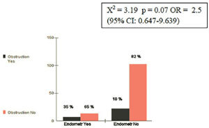
Figure 1. Endometriosis and Tubal Obstruction
DISCUSSION
Due to diagnostic difficulties and the different types, the literature has few high quality publications on endometriosis. Broeze et al. (2012) for example, state that this disease is a condition that may result in tubal pathology, but information on endometriosis was either not documented in the original databases or not reported in a standardized way, or was in sufficient detail. For these reasons the author could even included endometriosis as a clinical variable in a meta-analysis.
Removing endometriomas without hydrosalpinx or tubal obstruction remains controversial. Some authors state that this procedure did not improve the results of in vitro fertilization (Garcia-Velasco et al., 2004). Nevertheless assisted reproductive technology is better than surgery, and should be offered as a first-line treatment (Feinberg et al., 2008).
Osuga et al. (2008) published a case of hydrosalpinx and endometrioma without apparent infection. Salpingectomy was undertaken to improve the IVF-ET outcome. However most of the hydrosalpinx was an infection sequel, mainly Chlamydia. This publication did not find an association between endometriosis III and IV and tubal obstruction, thought the statistical test almost reached significance. Further studies with larger data sets are needed to check these results.
CONCLUSION
Although the OR was 2.5 (p=0.07) there was no significant difference between the groups with and without endometriosis III/IV. Further studies with larger samples are needed.
ACKNOWLEDGEMENT
The authors would like to thank the Human Reproduction Laboratory HC/GO to support this study. This study was financed in part by the Coordenação de Aperfeiçoamento de Pessoal de Nível Superior - Brasil (CAPES) - Finance Code 001.
REFERENCES
Bowman MC, Cooke ID. Comparison of fallopian tube intraluminal pathology as assessed by salpingoscopy with pelvic adhesions. Fertil SteriI. 1994;61:464-9.
Medline Crossref
Broeze KA, Opmeer BC, Coppus SF, Van Geloven N, Den Hartog JE, Land JA, Van der Linden PJ, Ng EH, Van der Steeg JW, Steures P, Van der Veen F, Mol BW. Integration of patient characteristics and the results of Chlamydia antibody testing and hysterosalpingography in the diagnosis of tubal pathology: an individual patient data meta-analysis. Hum Reprod. 2012;27:2979-90.
Medline Crossref
Feinberg EC, Levens ED, DeCherney AH. Infertility surgery is dead: only the obituary remains? Fertil Steril. 2008;89:232-6.
Medline Crossref
Garcia-Velasco JA, Mahutte NG, Corona J, Zúñiga V, Gilés J, Arici A, Pellicer A. Removal of endometriomas before in vitro fertilization does not improve fertility outcomes: a matched, case-control study. Fertil Steril. 2004;81:1194-7.
Medline Crossref
Goldberg JM, Bedaiwy MA. Recurrent umbilical endometriosis after laparoscopic treatment of minimal pelvic endometriosis: a case report. J Reprod Med. 2007;52:551-2.
Medline
Hickey M, Ballard K, Farquhar C. Endometriosis. BMJ. 2014;348:g1752.
Medline Crossref
Honoré GM, Holden AE, Schenken RS. Pathophysiology and management of proximal tubal blockage. Fertil Steril. 1999;71:785-95.
Medline Crossref
Lee A, Tran HT, Walters RF, Yee H, Rosenman K, Sanchez MR. Cutaneous umbilical endometriosis. Dermatol Online J. 2008;14:23.
Medline
Mahutte NG, Arici A. New advances in the understanding of endometriosis related infertility. J Reprod Immunol. 2002;55:73-83.
Medline Crossref
Marcoux S, Maheux R, Bérubé S. Laparoscopic surgery in infertile women with minimal or mild endometriosis. Canadian Collaborative Group on Endometriosis. N Engl J Med. 1997;337:217-22.
Medline Crossref
Osuga Y, Koga K, Hirata T, Hiroi H, Taketani Y. A case of hydrosalpinx associated with the menstrual cycle. Fertil Steril. 2008;90:199.e9-11.
Medline Crossref
Tanahatoe SJ, Hompes PG, Lambalk CB. Investigation of the infertile couple: should diagnostic laparoscopy be performed in the infertility work up programme in patients undergoing intrauterine insemination? Hum Reprod. 2003;18:8-11.
Medline Crossref
Toya M, Saito H, Ohta N, Saito T, Kaneko T, Hiroi M. Moderate and severe endometriosis is associated with alterations in the cell cycle of granulosa cells in patients undergoing in vitro fertilization and embryo transfer. Fertil Steril . 2000;73:344-50.
Medline Crossref