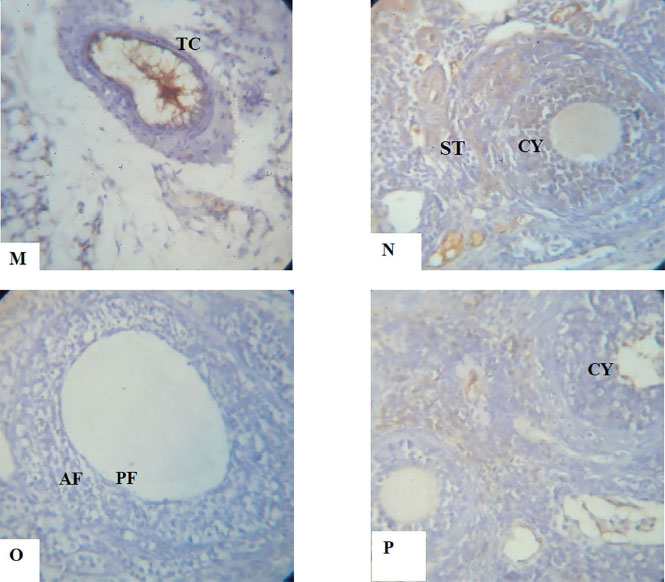
PLATES M-P. Show the immunohistochemical expression of the apoptotic protein Bax, anti-apoptotic protein Bcl2 and Adhesion molecule E-Cadherin photomicrograph sections of ovaries from Dehydroepiandrosterone (DHEA) -exposed rats and Controls following a 15-day treatment with aspirin using X40 magnifications.
M. Photomicrograph section of ovary from the Control group showing normal expression of E-cadherin which was largely confined to areas of the interstitium, theca and surface epithelium.
N. Photomicrograph section of ovary from DHEA-exposed rats, showing an increased number of cystic follicles (CY). Expression of E-Cadherin, which was largely confined to areas of the stroma, indicating a reduction in follicle growth.
O. Photomicrograph section of an ovary from Aspirin-exposed rats showing normal preantral and antral follicles. The granulosa cells are also normal with well-expressed E-Cadherin, indicating follicle growth and development.
P. Photomicrograph section of ovaries from Aspirin- and DHEA- exposed rats, showing a decreased number of cystic follicles. Expression of E-Cadherin was decreased in preantral and antral follicles, but well expressed in the granulosa cells. This indicates a significant improvement in ovary growth pattern.