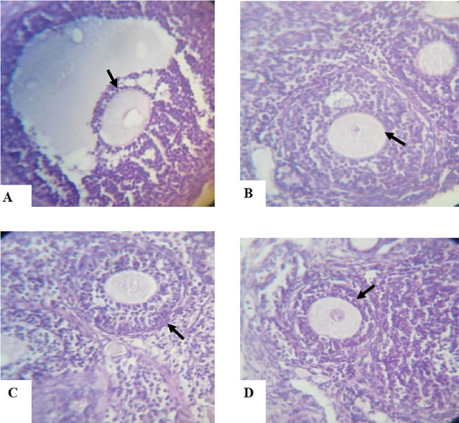
PLATES A–D. Histological sections of ovaries from Dehydroepiandrosterone (DHEA)-exposed rats and Controls following a 15-day treatment with aspirin using hematoxylin and eosin stain (H&E) X40 magnification.
A. Photomicrograph section of a Control-Group ovary showing normal follicle of different stages with well-organized surface epithelium, but without cystic follicle. The stromal cells of the ovarian follicles and corpus luteum are also well developed.
B. Photomicrograph section of ovary from DHEA-exposed rats showing an increased number of cystic follicles with a thicker theca cell layer as indicated by the arrow, and a marked higher level of collagen, especially in the region around the follicle. There is absence of corpora lutea and antral follicles.
C. Photomicrograph section of ovary from Aspirin-exposed rats showing normal histo-architecture of the ovary. The interstitial tissues are also normal.
D. Photomicrograph section of ovary from Aspirin- and DHEA-exposed rats, showing a decreased number of cystic follicles with a thinner theca cell layer and a reduced level of collagen, especially in the region around the follicle.