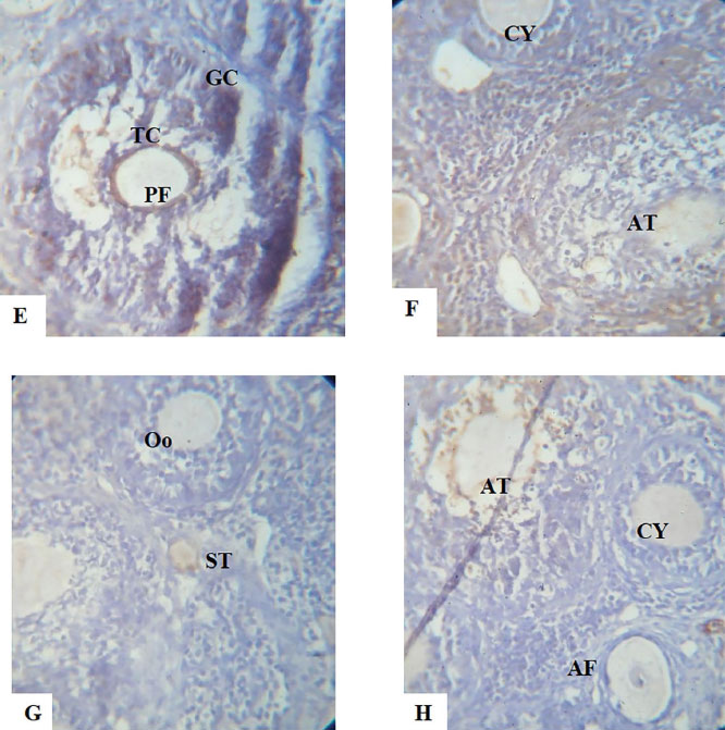
PLATES E-H. Show the immunohistochemical expression of the apoptotic protein Bax, anti-apoptotic protein Bcl2 and Adhesion molecule E-Cadherin photomicrograph sections of ovaries from Dehydroepiandrosterone (DHEA) -exposed rats and Controls following a 15-day treatment with aspirin using X40 magnifications.
E. Ovary photomicrograph section of the Control group showing normal preantral follicles (PF), granulosa cells (GC) and theca cells (TC). The Bax expression is mainly located in the granulosa cells of antral follicles.
F. Photomicrograph section of ovary from DHEA-exposed rats showing an increased number of cystic follicles (CY) and atretic follicles (AT), the Bax expression is higher in preantral and antral follicles, indicating a polycystic condition.
G. Photomicrograph section of ovary from Aspirin-exposed rats showing normal preantral, antral follicle and stroma (ST). The granulosa cells and Oocytes (Oo) are also normal.
H. Photomicrograph section of ovaries from Aspirin- and DHEA- exposed rats showing a decreased number of cystic follicles. The Bax expression is decreased in preantral and antral follicles (AF). This indicates improvements in the ovary’s cyto-architecture.