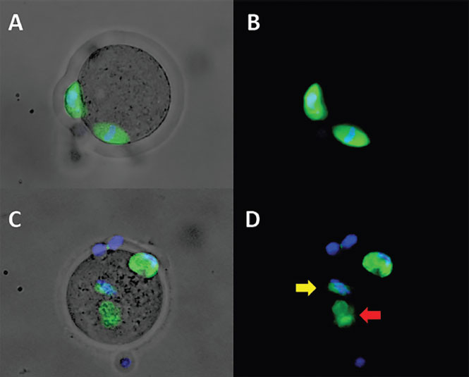
Figure 3. Representative immunofluorescence images of mouse oocytes stained for spindle and chromosomesA & B) MII oocyte, exposed to FF of oocyte donor, with normal spindle morphology and chromosomes correctly positioned in the equator. C & D) MII oocyte, exposed to FF of patient with endometriosis, showing abnormal spindle morphology (red arrow) and chromosomes localized apart from the spindle (yellow arrow).A & C) Composed image of transmitted light + spindle & chromosomal staining.B & D) Composed image of spindle & chromosomal staining.