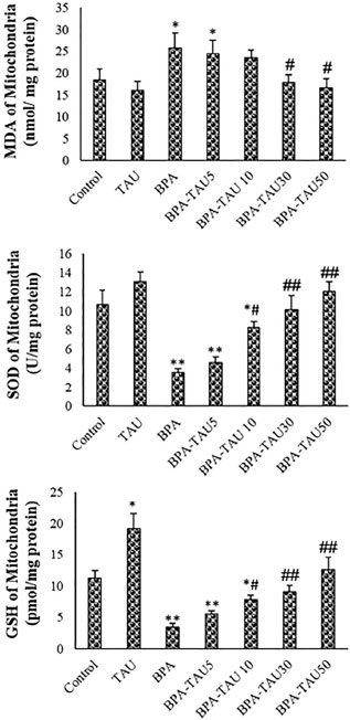
Figure 4. MDA, SOD and GSH levels of mice spermatozoa. The mean ± standard deviations are shown (n=6).MDA, SOD and GSH levels of the isolated mitochondria. The mean ± standard deviations are shown (n=6).* and # symbols show a comparison of the control and BPA groups, respectively.