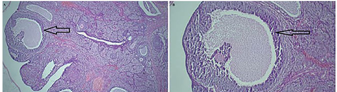
Figure 1. Light photomicrographs of an ovary in the control group. A. The structure of the ovarian tissue is normal with healthy growing follicles (H&E, ×40) and B. (H&E, ×100). Arrow: secondry follicle.
Figure 1. Light photomicrographs of an ovary in the control group. A. The structure of the ovarian tissue
is normal with healthy growing follicles (H&E, ×40) and B. (H&E, ×100). Arrow: secondry follicle.