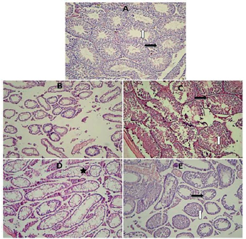
Figure 2. (A) A section from control group showing normal histological findings of preserved spermatogenesis. (B) This section of testis is from the busulfan group, showing damaged seminiferous tubules and interstitial tissue. (C) A section from the melatonin group showing preserved spermatogenesis up to the level of spermatocyte and Leydig cells. (D) A section from the melatonin group showing damaged and vacuolated tubules. (F) A section from the ozone group showing normal seminiferous tubules and interstitial tissue (white Arrows are showing spermatogenesis; black arrows are showing interstitial tissue with Leydig cells and Stars are showing vacuolated tubules. Hematoxylin and Eosin stain ×20.