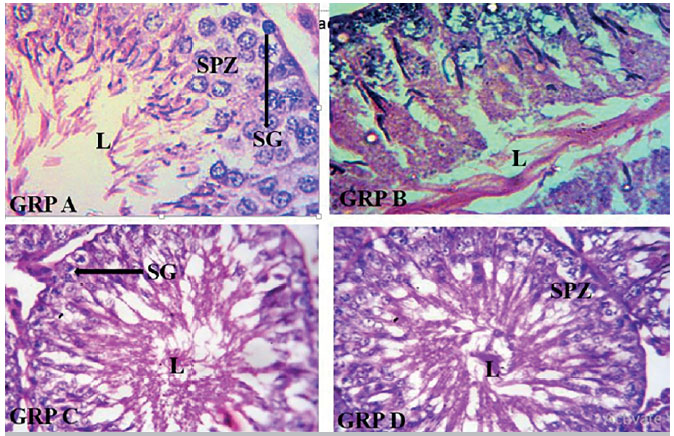
Plate 1. Photomicrograph of testes showing: Group A with normal histological architecture characterized by typically organized layers of spermatogenic cells; Group B with distorted tubular architecture and disorganization of the spermatogenic cells in seminiferous tubules and major pathological changes in the lumen (L); Group C with restored microarchitecture of the testicular morphology, distribution of epithelial lining, partially restored lumen (L); Group D with restored microarchitecture of the testicular morphology, distribution of epithelial lining, partially restored lumen (L). Stains: Hematoxylin and Eosin (H&E). Magnification X100.