JBRA Assist. Reprod. 2021;25(1):122-127
ORIGINAL PAPER
doi: 10.5935/1518-0557.20200069
Monozygotic twinning following embryo biopsy at the blastocyst stage
1Reproductive Medicine. Instituto Bernabeu, Alicante, Spain
CONFLICT OF INTEREST
The authors certify that they have NO affiliations with or involvement in any
organization or entity with any financial interest (such as honoraria;
educational grants; participation in speakers’ bureaus; membership,
employment, consultancies, stock ownership, or other equity interest; and
expert testimony or patent-licensing arrangements), or non-financial
interest (such as personal or professional relationships, affiliations,
knowledge or beliefs) in the subject matter or materials discussed in this
manuscript. No external funding was sought or obtained for this study
ABSTRACT
Objective: Monozygotic twinning incidence following preimplantation genetic testing in
embryos at cleavage-stage does not appear to increase; however, data
regarding the possible impact of the blastocyst-stage preimplantation
genetic testing is lacking. We compared the incidence of monozygotic
twinning in preimplantation genetic testing cycles performed at the
blastocyst-stage, versus cycles without PGT, following
single embryo transfer.
Methods: In this retrospective cohort study, we analyzed the incidence of twin
pregnancies in patients undergoing intracytoplasmic sperm injection and
blastocyst-preimplantation genetic testing (253 cycles), versus a period-matched control population of patients
undergoing intracytoplasmic sperm injection and single embryo transfer
without preimplantation genetic testing (606 cycles).
Results: The overall monozygotic twinning rate was 14/859 (1.6%) per clinical
pregnancy. The incidence of zygotic splitting following intracytoplasmic
sperm injection and preimplantation genetic testing was 3.5% (95% Confidence
interval 1.8%-6.6%) versus 0.8% (95% Confidence interval
0.3%-1.9%) following intracytoplasmic sperm injection without
preimplantation sperm injection. After adjusting for potential confounders,
preimplantation genetic testing cycles were associated with an increase in
the incidence of monozygotic twinning when compared to cycles without embryo
biopsy (Odd ratio 3.44, 95% Confidence interval 1.05-11.27, p=0.041).
Conclusions: Our findings indicate that embryo biopsy for preimplantation genetic testing
performed at the blastocyst stage is associated to an increase in the
incidence of monozygotic twinning. Further validation in larger sample size
studies is warranted. Patients undergoing preimplantation genetic testing
must receive proper counselling about the potential risks of the
technique.
Keywords: monozygotic twinning, embryo biopsy, blastocyst, preimplantation genetic testing
INTRODUCTION
Monozygotic twinning (MZT) (the splitting of a single fertilized oocyte into two or
more fetuses) occurs in 0.42% of spontaneous pregnancies (Bulmer, 1970). Monozygotic twin pregnancies are associated with
an increased risk of maternal and fetal complications, such as fetal growth
restriction, preterm delivery and perinatal mortality (Derom et al., 1987; Malone, 2003). The exact origin of MZT in spontaneous
pregnancies remains largely unknown and with the advent of assisted reproduction
techniques (ART), several publications have raised concerns regarding the potential
for an increased rate of MZT following ART (Abusheikha et al., 2000; Schachter et al., 2001). Recently, 1.5%
monozygotic twin live birth rate following ART was reported (Mateizel et al., 2016), which appears
concordant with the results from a large registered ART data analysis reporting a
prevalence of 1.36% of multiple pregnancies with zygotic splitting even after single
embryo transfer (Ikemoto et al.,
2018). Additional publications have investigated different ART parameters
and their putative involvement in embryo splitting and their potential association
with MZT. This includes: maternal age (Ikemoto et al., 2018), ovulation induction (Derom et al., 1987), embryo
culture conditions (Edwards et al.,
1986), changes to the zona pellucida (ZP) from ICSI and/or assisted
hatching (AH) (Hershlag et al.,
1999), cryopreservation and prolonged embryo culture up to blastocyst
stage (Nakasuji et al.,
2014). Taken all together, the body of available evidence suggests an (almost
uniform) association between blastocyst transfer and a potential risk of increased
MZT; whereas frozen embryo transfer (FET) appears associated with a lower MZT rate.
Other ART parameters appear equivocal and warrant future studies (Mateizel et al., 2016; Ikemoto et al., 2018).
Recently, following a revision in ART terminology (Zegers-Hochschild et al., 2017), the term
“preimplantation genetic testing” (PGT) was introduced, to describe the test
performed to analyze the DNA from oocytes (polar bodies) or embryos (cleavage stage
or blastocyst) for HLA typing or for determining genetic abnormalities. Publications
on the incidence of MZT after the use of PGT in ART cycles are scarce. At least
theoretically, the incidence of MZT in PGT cycles must be potentially increased
because of different mechanisms, including: intentional holes in the ZP and cell
manipulation for biopsy. To the best of our knowledge, the only available
publication on the topic reported that the incidence of MZT did not increase in PGT
cycles compared with regular intracytoplasmic sperm injection with blastocyst
transfer cycles (Verpoest et al.,
2009), this publication included cleavage-stage embryo biopsies
exclusively. More recently, cleavage-stage embryo biopsies have been largely
replaced by trophectoderm (TE) biopsies. Blastocyst biopsy provides more cells and
is performed at an embryonic stage more amenable to genetic analyses, and less
sensitive to possible damage (Coll et
al., 2018). We lack reports on the incidence of MZT after
the use of ART in combination with PGT performed at the blastocyst stage.
The aim of this study was to assess the incidence of MZT pregnancies in a cohort of
consecutive PGT treatment cycles performed at the blastocyst stage, followed by
single blastocyst embryo transfer, in comparison with a period-matched cohort of
consecutive ICSI cycles with single embryo transfer (SET) at the same center.
MATERIAL AND METHODS
In this retrospective comparative cohort study, we analyzed the incidence of MZT
pregnancies in a cohort of consecutive ICSI treatment cycles combined with PGT by TE
biopsy and single blastocyst embryo transfer (study group) and compared the results
with a control cohort of consecutive ICSI cycles and SET without embryo biopsy
(control group). The study was conducted at a single private center for reproductive
medicine and included data from January 2014 to December 2017.
Study and control groups
ART cycles included own-eggs and donor-eggs cycles. Specific characteristics of
the ovarian stimulation process were not included for analysis but there were no
differences in laboratory conditions vis-à-vis procedures between control and
study groups, except for the AH (on day-3 of embryo development) and TE biopsy
procedure for PGT in the study group. Once collected, the oocytes were incubated
in a culture medium (Global Total for Fertilization,
LifeGlobal®) until denudation for subsequent ICSI (usually
two hours later). In the study group, ICSI was performed in order to prevent
contamination with residual sperm DNA (Liebaers et al., 1998). The details of the ICSI procedure
have been described previously (Lledó et
al., 2006). Fertilization was assessed 16-18 hours after
ICSI and the embryos obtained remained in one-step culture medium (Global Total,
LifeGlobal®). Further development was evaluated in the
morning of day 3 when laser-AH (Lykos®, Hamilton
Thorne®) was performed as previously described (Lledó et al., 2006). At
the blastocyst stage, the embryos were classified according to morphologic
characteristics by ASEBIR (2015).
Laser-assisted biopsy and aspiration was applied consistently to remove five to
eight TE cells.
Within the study group, depending on clinical decision, biopsied embryos were
either selected for fresh transfer the day after biopsy (study-fresh group),
vitrifying supernumerary euploid embryos, or for elective complete vitrification
(study-frozen group) and deferred embryo transfer. Genetic analysis was
performed using array-CGH (which provides results within 24 hours) in the fresh
group, whereas Next Generation Sequencing (NGS) was used in the frozen group.
Similarly, in the control group and depending upon clinical decision, embryos
were selected either for fresh transfer (control-fresh group) plus vitrification
of surplus embryos or for elective complete vitrification (control-frozen group)
for deferred embryo transfer. A single embryo transfer was performed in all
cases; cleavage or blastocyst stage transfer was allowed in the control
group.
Endometrial preparation in frozen-thawed embryo transfer (FET) artificial cycles
(exogenous estrogen administration plus timely addition of progesterone) as well
as natural cycle endometrial preparation used as per clinical decision. Specific
characteristics of the endometrial preparation process were not included in the
analysis.
MZT diagnosis
A clinical pregnancy was defined as one or more gestational sacs seen via
transvaginal ultrasound scan at least 5 weeks after embryo transfer. MZT was
identified at first ultrasound when the number of fetal heartbeats exceeded the
number of gestational sacs, or when the number of sacs exceeded the number of
embryos transferred.
Statistical analysis
For the statistical analysis of patients and cycle characteristics, continuous
variables were presented as number of cases, mean and typical deviation. A p=0.05 was considered statistically significant after
performing a t-student test. Categorical variables were presented as number of
cases, percentage and odds ratio (OR). Pearson's Chi-square test (univariate)
and binary logistic regression (multivariate for confounding factors) were used
to analyze association of specific ART features and MZT. A p-value was considered significant if <0.05. The statistical
analysis was performed using the SPSS 20.0 software (SPSS, Chicago, IL,
USA).
RESULTS
Overall, we had 859 clinical pregnancies after SET during the study period. A total
606 cycles corresponded to ICSI and single embryo transfer without PGT and 253
cycles to patients undergoing ICSI and PGT (for various indications) with single
blastocyst transfer. Of these, 14 resulted in twin pregnancies (global MZT rate 1.6%
per clinical pregnancy). Globally, 93.3% of the cycles included embryo transfer at
the blastocyst stage. Table 1 depicts the
distribution of the baseline cycle characteristics within the MZT and singleton
groups. Monozygotic splitting occurred significantly more frequently
(p=0.004) in PGT cycles. Frozen embryo transfer cycles were
found more frequently associated with MZT without reaching statistical significance
(p=0.06).
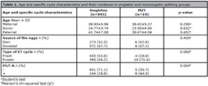
Table 1. Age and specific cycle characteristics and their incidence in singleton and monozygotic splitting groups
Table 2 shows that the incidence of zygotic splitting following PGT was 3.5% (95% CI 1.8% - 6.6%) versus 0.8% (95% CI 0.3% - 1.9%) following ICSI (without embryo biopsy).
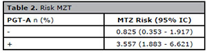
Table 2. Risk MZT
Table 3 presents a logistic regression analysis evaluating the effect of age and specific cycle parameters over the incidence of MZT. In the univariate analysis, cycles using donated eggs were more associated with a significant increased risk in monozygotic splitting when compared to own-eggs ICSI cycles (OR 5.01, 95% CI 1.1 - 22.69). The transfer of a frozen embryo also resulted in a significantly increased risk for MZT when compared to a fresh embryo in the univariate analysis (OR 4.12, 95% CI 1.04 - 16.18). After adjusting for these potential confounders (source of eggs and fresh/frozen cycles), the PGT procedure was associated with a significant increase in the incidence of MZT when compared to a non-biopsied embryo (OR 3.44, 95% CI 1.05-11.27, p=0.041).
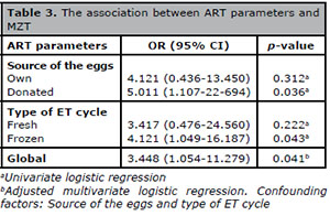
Table 3. The association between ART parameters and
MZT
Table 4 shows a sub-analysis considering blastocyst-stage transfers only in the control group. The frequency of monozygotic splitting remains statistically significant (p=0.006) in PGT cycles as occurred in the overall analysis (Table 1). However, after adjusting for the described potential confounders (source of the eggs and type of ET cycle), the PGT group was more frequently associated with MZT, but without reaching statistical significance (p=0.066).
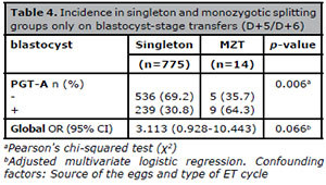
Table 4. Incidence in singleton and monozygotic splitting
groups only on blastocyst-stage transfers (D+5/D+6)
DISCUSSION
To the best of our knowledge, this is the first study focused on exploring the
potential effect of the embryo biopsy procedure at the blastocyst stage vis-a-vis
the incidence of monozygotic twin gestations. Notwithstanding the weaknesses
associated to its retrospective design (including residual confounding) and limited
sample, still our findings can contribute to the body of (scarce) medical evidence
on the subject. We report an increased risk of monozygotic splitting with the PGT
embryo biopsy procedure when performed at blastocyst stage in ART cycles.
The main strength of our study is the inclusion of cycles receiving only a single
embryo for transfer and a vast proportion of embryos transferred at the blastocyst
stage, thereby limiting the potential bias associated with the transfer of multiple
embryos at different embryonic stages.
Even after single embryo transfer, we had a 1.6% monozygotic twin rate, higher than
what is reported after spontaneous conception (0.40-0.45%; Aston et al., 2008), but similar to previous
reports in ART cycles (1.4%, Nakasuji et
al., 2014); 2.2%, Mateizel et al., 2016) including PGT only cycles (1.9%, Verpoest et al., 2009). Even
though the absolute numbers are quite small, these findings additionally confirm
that ART constitutes a risk factor for monozygotic pregnancies.
More specifically, within these SET cycles only, we considered exploring for certain
cycle parameters and their possible association with embryo splitting; the source of
the eggs: own vs donated (proxy for the potential impact of age), fresh vs frozen
embryo transfer cycle and TE biopsy for PGT. According to our data, donor-eggs
appear to be associated with an increased risk of MZT. This finding is concordant
with a previous publication (Luke et
al., 2014), but in disagreement with a previous
meta-analysis finding no association between the use of donor-eggs and the risk of
MZT (Busnelli et al., 2019),
among the possible explanations for these discrepancies are the number and stage of
the embryos transferred from oocyte-donors in other studies which (together with
other residual confounders) may introduce a source of bias. Interestingly, in the
case of own-eggs cycles, the meta-analysis by Busnelli et al. (2019) found that embryos derived from
younger oocytes (i.e., female age at time of retrieval <35 years) were
significantly more likely to result in an MZT pregnancy. Nonetheless, blastocyst
transfer was considered as a potential confounding factor, since it is well known
that embryos derived from young oocytes are more likely to be transferred at an
advanced blastocyst stage, finally concluding that age may not be an independent
risk factor for MZT but rather a proxy for blastocyst transfer (Busnelli et al., 2019).
However, in our study, the vast majority of embryos (93.3%) were transferred at the
blastocyst stage and still donor-eggs remained associated to an increased risk for
monozygotic splitting. As an additional note, one previous study has reported that
two MZT resulted from the transfer of embryos derived from a single oocyte donor's
retrieval and also another oocyte recipient who underwent a two-ET conceived
quadruplets (Knopman et al.,
2014). Further data is warranted to deepen in the exact etiology behind
the association of younger age and MZT, beyond the (probwably) valid but unspecific:
“healthier oocytes and superior reproductive potential” argument (Knopman et al., 2014).
MZT is a low rate event; thus, a high number of cases should be analyzed to find
significant differences. In terms of embryo stage upon ET, since the blastocyst
stage embryo transfer is highly predominant in our center, we expected a vast
majority of the control cases to fit into the blastocyst stage transfer category.
Indeed, the vast proportion of transfers in the control group (93.3%) were in the
blastocyst stage. To expand on the issue, we performed statistics including
blastocyst-stage transfers only day-5/day-6 (n=775), which resulted in similar
outcomes in the crude analyses, consistently showing an increase in the MZT rate
associated with the embryo biopsy procedure (Table
4). Nonetheless, -and presumably as a consequence of the reduction in
sample size-, the significance is lost when accounting for confounders in the data
analysis, even though a trend is evident. Taken all together, even acknowledging the
absence of statistical significance in the blastocyst-only sub-analysis, our data is
still concordant with an increased risk for MZT associated with the embryo biopsy
procedure.
Regarding fresh vs. frozen cycles, our study found that the transfer
of a frozen embryo was associated with an increased risk of MZT in the univariate
analysis. The same finding has been reported by several (Alikani et al., 2003; Knopman et al., 2014; Nakasuji et al., 2014; Ikemoto et al., 2018) but not all previous
publications (Mateizel et al.,
2016; Busnelli et al.,
2019) on the topic. It is worth noting that a significant proportion of
PGT cycles in our study were transferred in a frozen cycle (148/253, 58.5%). Hence,
the possibility exists that the frozen cycle may act as a confounding factor.
Consequently, frozen cycles might not be a true independent risk factor for MZT, but
rather a proxy for PGT impact. An important limitation of our observational study is
that data regarding frozen-warmed embryo transfer cycles did not include information
about specific endometrial preparation protocols, which may have influenced the
outcome (e.g., an endometrial preparation in a natural cycle may have contributed to
an “additional” natural conception). Therefore, a causal/casual relationship between
zygotic splitting and embryo-cryopreservation-frozen/thaw ART procedures might be
suggested, but cannot be demonstrated from our data.
Embryo PGT entails several invasive procedures and manipulations including breaks,
(laser-assisted) intentional holes in the ZP and TE cells biopsy, all of which could
potentially increase MZT rates (Verpoest et
al., 2009). According to our data, even after adjusting for
potential confounders, PGT cycles remain a significant factor associated with an
increased risk for monozygotic splitting compared to ICSI cycles. Our findings
contradict a previously reported data on the topic, which showed no increase in MZT
rate and PGT-A compared to regular ICSI after fresh blastocyst transfer (Verpoest et al., 2009). This
divergence may be attributed to several factors. First, the number of embryos for
transfer. In the study by Verpoest et
al. (2009), a mean of 1.6 embryos (in each arm) were
transferred and, -as acknowledged by authors-, chorionicity was not clearly
established in MZT pregnancies. This policy may have introduced a substantial bias
since a causal relationship between the higher number of embryos transferred and MZT
has been argued (Sills et al.,
2000). In our study, albeit chorionicity or DNA fingerprinting were also
not addressed, the inclusion of SET only cycles would have limited this potential
source of bias. Secondly, the inclusion of fresh-only cycles by Verpoest et al. (2009); as
previously discussed, almost 60% of PGT cycles in our study included frozen-thawed
cycles, which may have additionally contributed to the MZT outcome as suggested in
the univariate analysis. Specific changes in the ZP or TE cells during/after
vitrification in a blastocyst-stage biopsied embryo could be hypothesized as
potential additional factors associated with the increased risk of MZT in frozen
cycles.
This hypothesis warrants further investigations. Third, and of the utmost importance,
the difference in the embryo-stage when performing the biopsy. The study by Verpoest et al. (2009)
included cleavage-stage embryo biopsies, which is a less invasive approach if
compared with the TE blastocyst stage embryo biopsy. Additional to the procedures
for cleavage-stage biopsies, blastocyst biopsies add aspiration and laser-assisted
excision in combination with mechanical removal of TE cells by “pulling” or
“flicking”. A possible factor explaining the split of the inner mass cell (IMC)
during long culture up to the blastocyst stage for subsequent embryo biopsy is the
exposure of the embryo to low concentrations of Ca+2 in the culture
media. These low concentrations may promote less cells-adhesion and a “weakening” of
the inter-cellular junctions provoking the detachment of some cells from the IMC and
the formation of an extra embryonic pole in the TE (Milki et al., 2003). Therefore, it can be hypothesized
that the exposure of the embryo to low Ca+2 concentrations together with
the mechanical manipulation during embryo biopsy may act as added factors (chemical
and mechanical) favoring a later splitting of the ICM, resulting in MZT. While some
reports may support this hypothesis (Edwards et al., 1986; Van
Langendonckt et al., 2000; Yan et al., 2015), a recent retrospective
evaluation focused on PGT cycles (Gu et
al., 2018) taking into account the “hatching” status of the
blastocyst (partially and fully hatched blastocysts vs 8-shaped blastocysts with ICM
incarceration) before biopsy and the relation with embryo splitting, showed that ICM
incarceration in 8-shaped blastocysts did not increase MZT incidence. While some
differences in the protocol may explain this discrepancy, Gu et al. (2018) still reported an overall MZT
pregnancy rate per established clinical pregnancy of 2.8% (similar to our rate).
Unfortunately, the study did not include regular ICSI with blastocyst transfer for
comparison. In concordance with our study, the (additional) influence of AH in the
MZT rate could not be disentangled in the study by Gu et al. (2018), because all the embryos in the PGT
group received AH on day 3 of embryo development. But, as suggested by some
observations, even if the zona manipulation on cleavage-stage (i.e., AH) may be
involved in some cases of MZT, it is unlikely to be an exclusive mechanism (Gu et al., 2018). Nonetheless,
in the study by Verpoest et al.
(2009) cleavage stage embryo-biopsies did not show correlation with an
increase in MZT rate; in these regards, cleavage-stage biopsy could be inferred as a
proxy for AH (both procedures involves “making a hole” in the zona pellucida during
cleavage stage).
Also, some additional drawbacks from our study merits further analysis. MZT is -
fortunately- an infrequent event; indeed studies focused on rare phenomenon
constitute a challenge. Obviously, the results from our study, with a limited sample
size, should be further explored in large databases in order to reach robust
conclusions. However, due to the sharp increase in PGT cycles worldwide, even small
increases in incidences may translate into important crude numbers of this
potentially complex obstetric condition. The results from our study may inspire
future studies on the subject. The lack of confirmation of monozygocity at birth
constitutes another arguable limitation of the study; we believe that this approach
is only of marginal importance, and it is not crucial for the purpose of diagnosis
of am MZT (especially after SET policy); several publications on the topic have
clearly established that transvaginal ultrasound is highly accurate in diagnosing a
MZT pregnancy; moreover all cases in our study received one additional and dedicated
evaluation of the MZT status on the week after the initial visualization for
confirmation (weeks 6-7). Notwithstanding these potential limitations, still the
results found in our study can contribute to the body of medical evidence on the
subject.
CONCLUSION
Our findings indicate that embryo biopsy for PGT performed at the blastocyst stage is
associated to an increase in the incidence of MZT. Because of the increased risk
associated with MZT, this information could be useful to help identify potential
factors underlying the higher incidence of MZT in the context of ART, and to
identify strategies to reduce this incidence. Given the low event rates with
monozygotic twinning, our results warrant validation in larger sample size studies
required to provide higher statistical power. Until those studies become available,
patients undergoing PGT must receive proper counselling about the potential risks of
the technique.
REFERENCES
Abusheikha N, Salha O, Sharma V, Brinsden P. Monozygotic twinning
and IVF/ICSI treatment: a report of 11 cases and review of literature. Hum
Reprod Update. 2000;6:396-403.
Medline Crossref
Alikani M, Cekleniak NA, Walters E, Cohen J. Monozygotic twinning
following assisted conception: an analysis of 81 consecutive cases. Hum Reprod.
2003;18:1937-43.
Medline Crossref
ASEBIR - Asociación para el Estudio de la Biologia de la Reproduction. Criterios ASEBIR de Valoración Morfológica de Oocitos, Embriones Tempranos y Blastocistos Humanos. Cuadernos de Embriología Clínica [Internet]; 2015. Available at: https://asebir.com/cuadernos-asebir/criterios-asebir-de-valoracion-morfologica-de-oocitos-embriones-tempranos-y-blastocistos-humanos/
Aston KI, Peterson CM, Carrell DT. Monozygotic twinning associated
with assisted reproductive technologies: a review. Reproduction.
2008;136:377-86.
Medline Crossref
Bulmer MG. The biology of twinning in man. Oxford: Clarendon Press; 1970.
Busnelli A, Dallagiovanna C, Reschini M, Paffoni A, Fedele L,
Somigliana E. Risk factors for monozygotic twinning after in vitro
fertilization: a systematic review and meta-analysis. Fertil Steril.
2019;111:302-17.
Medline Crossref
Coll L, Parriego M, Boada M, Devesa M, Arroyo G, Rodríguez I,
Coroleu B, Vidal F, Veiga A. Transition from blastomere to trophectoderm biopsy:
comparing two preimplantation genetic testing for aneuploidies strategies.
Zygote. 2018;26:191-8.
Medline Crossref
Derom C, Vlietinck R, Derom R, Van den Berghe H, Thiery M. Increased
monozygotic twinning rate after ovulation induction. Lancet. 1987;1:1236-8.
Medline Crossref
Edwards RG, Mettler L, Walters DE. Identical twins and in vitro
fertilization. J In Vitro Fert Embryo Transf. 1986;3:114-7.
Medline Crossref
Gu YF, Zhou QW, Zhang SP, Lu CF, Gong F, Tan YQ, Lu GX, Lin G. Inner
cell mass incarceration in 8-shaped blastocysts does not increase monozygotic
twinning in preimplantation genetic diagnosis and screening patients. PLoS One.
2018;13:e0190776.
Medline Crossref
Hershlag A, Paine T, Cooper GW, Scholl GM, Rawlinson K, Kvapil G.
Monozygotic twinning associated with mechanical assisted hatching. Fertil
Steril. 1999;71:144-6.
Medline Crossref
Ikemoto Y, Kuroda K, Ochiai A, Yamashita S, Ikuma S, Nojiri S,
Itakura A, Takeda S. Prevalence and risk factors of zygotic splitting after 937
848 single embryo transfer cycles. Hum Reprod. 2018;33:1984-91.
Medline Crossref
Knopman JM, Krey LC, Oh C, Lee J, McCaffrey C, Noyes N. What makes
them split? Identifying risk factors that lead to monozygotic twins after in
vitro fertilization. Fertil Steril. 2014;102:82-9.
Medline Crossref
Liebaers I, Sermon K, Staessen C, Joris H, Lissens W, Van Assche E,
Nagy P, Bonduelle M, Vandervorst M, Devroey P, Van Steirteghem A. Clinical
experience with preimplantation genetic diagnosis and intracytoplasmic sperm
injection. Hum Reprod. 1998;13:186-95.
Medline Crossref
Lledó B, Ten J, Galán FM, Bernabeu R. Preimplantation genetic
diagnosis of Marfan syndrome using multiple displacement amplification. Fertil
Steril. 2006;86:949-55.
Medline Crossref
Luke B, Brown MB, Wantman E, Stern JE. Factors associated with
monozygosity in assisted reproductive technology pregnancies and the risk of
recurrence using linked cycles. Fertil Steril. 2014;101:683-9.
Medline Crossref
Malone FD. Monochorionic pregnancy--where have we been? Where are we
going? Am J Obstet Gynecol. 2003;189:1308-9.
Medline Crossref
Mateizel I, Santos-Ribeiro S, Done E, Van Landuyt L, Van de Velde H,
Tournaye H, Verheyen G. Do ARTs affect the incidence of monozygotic twinning?
Hum Reprod. 2016;31:2435-41.
Medline Crossref
Milki AA, Jun SH, Hinckley MD, Behr B, Giudice LC, Westphal LM.
Incidence of monozygotic twinning with blastocyst transfer compared to
cleavage-stage transfer. Fertil Steril. 2003;79:503-6.
Medline Crossref
Nakasuji T, Saito H, Araki R, Nakaza A, Nakashima A, Kuwahara A,
Ishihara O, Irahara M, Kubota T, Yoshimura Y, Sakumoto T. The incidence of
monozygotic twinning in assisted reproductive technology: analysis based on
results from the 2010 Japanese ART national registry. J Assist Reprod Genet.
2014;31:803-7.
Medline Crossref
Schachter M, Raziel A, Friedler S, Strassburger D, Bern O, Ron-El R.
Monozygotic twinning after assisted reproductive techniques: a phenomenon
independent of micromanipulation. Hum Reprod. 2001;16:1264-9.
Medline Crossref
Sills ES, Moomjy M, Zaninovic N, Veeck LL, McGee M, Palermo GD,
Rosenwaks Z. Human zona pellucida micromanipulation and monozygotic twinning
frequency after IVF. Hum Reprod. 2000;15:890-5.
Medline Crossref
Van Langendonckt A, Wyns C, Godin PA, Toussaint-Demylle D, Donnez J.
Atypical hatching of a human blastocyst leading to monozygotic twinning: a case
report. Fertil Steril. 2000;74:1047-50.
Medline Crossref
Verpoest W, Van Landuyt L, Desmyttere S, Cremers A, Devroey P,
Liebaers I. The incidence of monozygotic twinning following PGD is not
increased. Hum Reprod. 2009;24:2945-50.
Medline Crossref
Yan Z, Liang H, Deng L, Long H, Chen H, Chai W, Suo L, Xu C, Kuang
Y, Wu L, Lu S, Lyu Q. Eight-Shaped Hatching Increases the Risk of Inner Cell
Mass Splitting in Extended Mouse Embryo Culture. PLoS One. 2015;10:e0145172.
Medline Crossref
Zegers-Hochschild F, Adamson GD, Dyer S, Racowsky C, de Mouzon J,
Sokol R, Rienzi L, Sunde A, Schmidt L, Cooke ID, Simpson JL, van der Poel S. The
International Glossary on Infertility and Fertility Care, 2017. Fertil Steril.
2017;108:393-406.
Medline Crossref