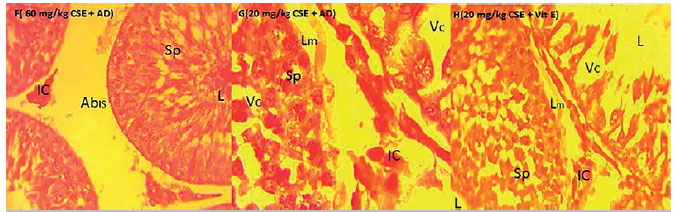
Plate F. Demonstrates testis of animal treated with 60mg/kg CSE+AD; abnormal widening of Interstitial spaces, Abis, however, all the stages of spermatogonia population expressed, SB-Spermatogonial, SASpermatogonia B, SE-Seminiferous Epithelium, IC-interstitial cells, SL-Sertoli cells. Indication of continuous spermatogenesis with the presences of spermatocytes in the lumen H&E, x400
Plate G. Demonstrates testis of animal treated with 20mg/kg CSE+AD; Reduced abnormal widening of interstitial paces, mild alteration in the all architecture seen in the loosening of the SE-Seminiferous Epithelium, reduced evidence of vacuolation; however, with spermatogonia regeneration and continuous spermatogenesis. H&E stain, X1000.
Plate H. Demonstrates testis of animals treated with 60mg/kg CSE+Vit. E; Spermatogonia population with evident of regeneration and a reduced vacuolation. Interstitial spaces intact with the Leydig cells and reduced loose basal seminiferous membrane with continuous spermatogenesis. H&Stain, x 400