JBRA Assist. Reprod. 2020;24(3):340-346
REVIEW ARTICLE
doi: 10.5935/1518-0557.20200046
The different tests for the diagnosis of COVID-19 - A review in Brazil so far
1Universidade Federal do ABC, Centro de Ciências Naturais e Humanas, São Bernardo do Campo – SP
2Igenomix, São Paulo – SP
The author has no conflict of interest to declare
ABSTRACT
SARS-CoV-2 is a novel virus from the coronavirus family that emerged in the end
of December 2019 in Wuhan, China. The virus is now widespread and causing the
current pandemic of COVID-19, a highly pathogenic viral pneumonia, commonly
presented with fever and cough, which frequently lead to lower respiratory tract
disease with poor clinical outcomes associated with older age and underlying
health conditions. Supportive care for patients is typically the standard
protocol because no specific effective antiviral therapies have been identified
so far. The current outbreak is challenging governments and health authorities
all over the world. In here we present a comparison among the current diagnostic
tools and kits being used to test Brazilian population.
Keywords: Immunochromatography, ELISA, PCR, diagnostic tool, COVID-19
Background
The coronavirus disease 2019 (COVID-19) epidemic started in December 2019, in Wuhan,
Hubei province, in China. It rapidly spread across China and other countries,
raising major global concerns (Tang et
al., 2020). Its etiological agent is the SARS-CoV-2 (Wu et al., 2020) also referred to as HCoV-19 (Jiang et al., 2020). According
to the latest update by the World Health Organization (WHO, 2020a) up to April 28,
2020 there were 2,959,929 confirmed cases with 202,733 deaths in 213 countries,
areas or territories so far.
The current COVID-19 outbreak is both similar and different to the prior SARS
(2002-2003) and MERS (2012-ongoing) outbreaks. SARS was initiated by zoonotic
transmission of a novel coronavirus (likely from bats via palm civets) in markets in
Guangdong province, China. MERS was also traced to zoonotic transmission of a novel
coronavirus (likely from bats via dromedary camels) in Saudi Arabia. All three viral
infections commonly presented with fever and cough, which frequently lead to lower
respiratory tract disease with poor clinical outcomes associated with older age and
underlying health conditions (Wu & McGoogan,
2020).
The treatment of COVID-19 is supportive. To date, no vaccine, antiviral or other
specific treatment is available, however, there are several studies in progress
(Wu & McGoogan, 2020). Also, it is
not known whether infectiousness starts before onset of symptoms. The incubation
period for COVID-19 is about 5-6 days (Li et
al., 2020a). Combining this time with a similar length
serial interval suggests there might be considerable presymptomatic infectiousness
(Anderson et al., 2020).
So far there have been few clinical studies to measure COVID-19 viremia and how it
changes over time in individuals (Anderson et al., 2020). In one
study of 17 patients diagnosed with COVID-19, peak viremia seems to be at the end of
the incubation period (Zou et al.,
2020), pointing to the possibility that viremia might be high enough to
trigger transmission for 1-2 days before onset of symptoms.
Diagnostic tests for COVID-19 have stood out in the current coronavirus pandemic as
an essential tool for tracking the spread of the disease. The genetic sequence of
the 2019 novel coronavirus enabled the rapid development of diagnostics tests
specific for SARS-CoV-2 (Wang et
al., 2020). Since there is a wide range of diagnostic tests
commercially available for SARSCoV-2, in this review we present a comparison among
of all them and the techniques used to test Brazilian population.
The Brazilian perspective and diagnostic tests available
In Brazil, the first case of COVID-19 was confirmed on February 26, 2020 by the
Ministry of Health. A 61-year-old man was admitted to a private hospital with a
history of travel to Italy, but he was already at home when he presented the
symptoms. Since then, on April 25, 2020, 58,509 cases have been confirmed, most
of them in the state of São Paulo. Figure 1 illustrates the number of cases per state in Brazil.
Brazilian Health Regulatory Agency (Anvisa) published the Resolution (RDC
348/2020), which established extraordinary and temporary rules to speed up the
evaluation of new products by prioritizing the analysis of test registration
requests for detection of the new coronavirus (SARSCoV-2). The idea is not to
evaluate and approve products automatically, as sanitary rigor must always
exist, but rather to speed up the process. The measure is part of the strategic
actions to enable products that can be used to face the COVID-19 pandemic.
Anvisa’s role is to promote the protection of the population’s health by
executing sanitary control of the production, marketing and use of products and
services subject to health regulation, including related environments,
processes, ingredients and technologies, as well as the control in ports,
airports and borders. On March 19, the Brazilian Health Regulatory Agency
approved the first eight rapid tests for the diagnosis of COVID-19. At the date
of the review, 39 tests have been approved by Anvisa so far. Of the 39 approved,
21 are rapid tests. There are tests that use blood, serum or plasma and others
that need samples of secretions collected from the airways, such as nasopharynx
(nose) and oropharynx (throat). Table 1 and 2 summarizes all kits and tests
registered in Brazil. Data regarding accuracy have been extracted from manual
instructions available at Anvisa website.
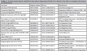
Table 1. All tests authorized by ANVISA to be used in diagnosing COVID-19 in
Brazil so far (NA no available information at 20/04/2020)
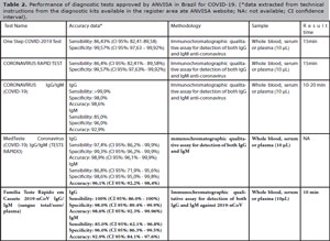
Table 2. Performance of diagnostic tests approved by ANVISA in Brazil for
COVID-19. (*data extracted from technical instructions from the
diagnostic kits available in the register area ate ANVISA website; NA:
not available; CI confidence interval).
Rapid Test or Point of care testing (POCT)
Most rapid tests use colloidal gold particles in a technique known as
immunochromatography, also called lateral flow immunoassay, a type of sandwich
assay that relies on a pair of antibodies used to recognize two independent
epitopes of a protein, and therefore it can achieve high specificity (Zhou et al., 2012).
Lateral flow assays only require the application of a sample (sometimes followed
by the application of a buffer solution) and can yield a result within 5-15
minutes (O’Farrell, 2009). This kind of
test is being used for pregnancy (Puertas et al., 2010), HIV (Granade et al., 2010), bacterial
infections (Huang, 2007) , drugs of abuse
(Gonzalez et al.,
2011), food contaminants (No et al., 2007) and dengue virus (Cuzzubbo et al., 2001), and
many tests are commercially available (Zhou et al., 2012). Lateral flow assays are also
being developed for global health applications, where devices that are
inexpensive and easy-to-use are required. However, lateral flow assays are
generally not quantitative and often only give a yes/no answer (Zhou et
al., 2012).
A typical immunochromatographic strip is composed by a sample-loading pad (O’Farrell, 2009), a glass fibre pad with
detection antibody (dAb) conjugated to gold nanoparticles (AuNPs) or latex beads
(Xu et al., 2007), a
nitrocellulose or polyvinylidene fluoride membrane with pre-immobilized capture
antibody (cAb), a control antibody for test validation (Puertas et al., 2010) and an absorbent pad
used as capillary pump to draw the sample solution (O’Farrell, 2009).
To perform a lateral flow, assay the sample containing the target analyte
(antigen) is loaded on the sample pad and flows through the membrane by
capillary effects. The liquid first dissolves the dAb-AuNP conjugates and the
antigen binds to the dAb. As the antigen-dAb pair flows through the capture
zone, the cAb will capture the labelled antigen. Further downstream, the unbound
dAb-AuNP reacts with the control antibody, which binds specifically to dAb
irrespective of the antigen. Both the capture and control lines may become
visible due to the accumulation of the AuNPs that produce collective plasmonic
effects and result in a red colour (Xu et al., 2007). The colour on the control line
indicates the test is valid, and the colour on the capture line suggests the
presence of target analyte in the sample solution (Zhou et al., 2012).
For COVID-19 the tests have been developed with AuNPs conjugated with
recombinant-anti-COVID-19 antigens. The sample (whole blood, serum or plasma) is
added to the pad and, the antibodies against COVID-19 present in the sample,
interact with the AuNPs and run through the membrane. When in contact with the
test regions these gold-antibodies conjugated are immobilized and a colour line
appears on the strip. The presence of the line indicates a positive result. The
absence indicates a negative result. There is also a control line, as an
indicative that the test is valid. Independent on the result the test is only
valid if the control line appears. Figure 2 is an example of how this technology works.
In this context many companies and research groups developed rapid lateral flow
immunoassay tests for testing specific antibodies of SARS-CoV-2 in patient blood
for being a rapid, simple, highly sensitive diagnosis (Li et al., 2020b).
One of the first rapid tests (lateral flow immunoassay) for SARS-CoV-2 IgG and
IgM immune responses was developed by professor´s Feng Ye group at the National
Clinical Research Centre for Respiratory Disease in Guangzhou, China. The
clinical efficiency of the tests was validated by collecting blood samples from
397 PCR confirmed COVID-19 patients and 128 negative patients at 8 different
clinical sites. The overall testing sensitivity was 88.66% and specificity was
90.63% (Li et al.,
2020b). This combined test is being designed and manufactured by Jiangsu
Medomics Medical Technologies (Nanjing, China).
From the 39 tests approved 21 are immunochromatographic tests to evaluate the
presence of IgG and IgM antibodies against SARS-CoV-2. Sample material for those
tests are whole blood, serum or plasma and the time for yielding a result varies
from 10-20 minutes. Specificity to IgM antibodies was 94-98% according to the
manufacturer while for the IgG was 97-98%. The sensibility for the IgM
antibodies was 85-90% and for IgG 95-100%. Three tests - One Step COVID-2019
Test (Celer Biotecnologia S/A), CORONAVÍRUS RAPID TEST (Biocon Diagnósicos) and
DPP® COVID-19 IgM/IgG System (Orangelife) had no available
data regarding sensibility and specificity for IgG and IgM.
The manufacturers of the tests CORONAVÍRUS IgG/IgM (COVID-19) and 2019-nCoV
IgG/IgM Teste Rápido em Cassete reported that hematocrit levels may affect
results. The hematocrit level should be between 25% - 65% to yield accurate
results. The manufacturer of MedTeste Coronavírus (COVID-19) IgG/IgM informed
that results of immunocompromised patients should be interpreted with caution.
The manufacturer of the test Anti COVID-19 IgG/IgM Rapid Test declared that no
false results were observed in positive sample for the pathogens: influenza A,
influenza BRSV, adenovirus, HBsAg syphilis, Helicobacter pylori, HIV and
HCV.
In general, this kind of assay shows some advantages such as: it is an
established mature technology; relative ease of manufacture since equipment and
processes are already developed and available; easily scalable to high volume
production, stable, since shelf-lives varies from 12 to 24 months often without
refrigeration; ease of use; relatively low cost and short timeline for
development and approval (O’Farrell,
2009). However, we have found relatively little current information
reporting the diagnostic performance of these POC devices using clinical samples
taken from community settings. Relevant data may still be under collection in
ongoing studies or may not be published publically.
ELISA
The enzyme immunoassays (EIA) and enzyme-linked immunosorbent assays (ELISA) are
both widely used as diagnostic tools for the detection and quantification of
specific antigens or antibodies in a given sample (Gan & Patel, 2013). Both techniques share similar basic principles
and are derived from the radioimmunoassay (RIA). RIA was first described by Berson
and Yalow, for which Yalow was awarded the Nobel Prize in 1977, to measure
endogenous plasma insulin (Yalow & Berson, 1996). RIA was then developed into a
novel technique to detect and measure biological molecules present in exceedingly
small quantities, paving the way for more analysis and detection of countless other
biological molecules, including hormones, peptides and proteins. Because of the
safety concerns regarding its use of radioactivity, RIA assays were modified by
replacing the radioisotope with an enzyme, thus creating the modern-day EIA and
ELISA (Gan & Patel, 2013).
Both assays use the basic immunology concept of an antigen binding to its specific
antibody, which allows detection of small quantities of antigens such as proteins,
peptides, hormones or even antibodies in a fluid sample. Those assays utilize
enzyme-labelled antigens and antibodies to detect biological molecules; the most
used enzymes being alkaline phosphatase (EC 3.1.3.1) and glucose oxidase (E.C.
1.1.3.4). The antigen in fluid phase is immobilized, usually in a 96-well microtiter
plate. The antigen can bind to a specific antibody, which is itself subsequently
detected by a secondary, enzyme-coupled antibody. A chromogenic substrate for the
enzyme yields a visible colour change or fluorescence, indicating then the presence
of the antigen. Quantitative or qualitative measures can be assessed based on such
colorimetric reading. (Gan & Patel,
2013).
Although ELISA methodology could help track antigen exposure, it has some
limitations: the enzyme-mediated colour change will react indefinitely. Over a
sufficient long period of time, the colour strength will inaccurately reflect the
amount of primary antibody present, yielding false-positive results; to detect a
given antibody or antigen, a known reciprocal antigen or antibody must be generated
and, nonspecific binding of the antibody or antigen to the plate will lead to a
falsely high-positive result (Gan & Patel,
2013).
Polymerase Chain Reaction
In acute respiratory infection, RT-PCR is routinely used to detect causative
viruses from respiratory secretions. PCR is an enzyme-driven process for
replicating DNA in vitro. PCR can produce enough amounts of DNA so that
pathogens can be detected and identified. Because each pathogen has a unique
complement of DNA or RNA, those molecules can function as a molecular
fingerprint to help identify what is the organism causing one disease. In this
technique a segment of DNA is copied in vitro by using a thermostable DNA
polymerase enzyme in the presence of buffer, magnesium, deoxyribonucleoside
triphosphates and primers. Oligonucleotide primers complementary to regions on
the coding and the noncoding strand of the DNA template are responsible for
specificity in the reaction, determining which region of the DNA becomes
amplified. As the primers anneal to their complementary regions of DNA, DNA
polymerases attach to the primer-template complexes and extend the DNA strands,
producing a copy of the DNA. Each copy may then serve as another template for
further amplification. Multiple rounds of heating and cooling of the reaction
mixture in a thermal cycler produce rounds of melting of the double-stranded
DNA, annealing of primer to single-stranded templates, and extension of DNA
strands, to produce a logarithmic increase in DNA. In the ideal scenario, the
primers chosen in the PCR are specific for a pathogen gene, and hence do not
amplify nonspecific targets such as human genes. Theoretically, one could start
with a single copy of the target pathogen gene present in the reaction and
generate billions of copies of DNA from that gene (Fredricks & Relman, 1999).
There are several approaches for using PCR to detect pathogen DNA, the simplest
one being specific PCR where the primers are designed to attach to complementary
regions of a DNA target (specific to the pathogen that is being assayed).
Broad-range PCR attempt to detect a broader group of organisms by designing
primers that are complementary to conserved regions of a particular gene that
are shared by a given taxonomic group (Relman,
1998). Another variation is multiplexing, in which multiple specific
PCR assays are run simultaneously in the same reaction tube test for multiple
different DNA templates. In multiplex PCR several sets of primers are added to
the reaction in order to generate several different PCR products. In this case
postamplification methods are needed to determine which organism is represented
in a positive reaction (Fredricks & Relman,
1999).
PCR is possibly the most quintessential molecular method yet developed.
Real-time quantitative PCR (qPCR) revolutionized clinical application of PCR
partly because it automated analysis by removing the need for postreaction
manipulation (Huggett et al.,
2015). qPCR is over 20 year old (Higuchi et al., 1992), but it has only really been
applied clinically in areas which alternatives are not practically possible,
such as monitoring treatment in diseases like chronic-phase chronic myeloid
leukaemia (Cross et al.,
2012) or for some key blood borne viruses (Fryer et al., 2008).
Three tests use RT-PCR as the core technology to detect SARS-CoV-2 in samples of
the airways of the patients, targeting the conserved genes ORF1ab and N. Probes
with fluorescent reporter dye are used to make the detection. Positive results
are indicative of the presence of RNA of the virus, but a clinical correlation
with patient´s history is necessary. Tests are highly sensitive. These data were
provided by Secretaria de Ciência, Tecnologia, Inovação e Insumos Estratégicos
em Saúde - SCTIE (2020).
When should we use all the tests?
On 2 March 2020, WHO released an interim guidance regarding laboratory testing
for COVID-19 in suspected human cases recommending that tests should be based on
clinical and epidemiological factor and linked to assessment of the likelihood
of infection. But is also recommended PCR testing of asymptomatic or mildly
symptomatic cases (WHO, 2020b). We should keep in mind that the gold standard
for diagnosing COVID-19, as referred by WHO, are nucleic acid amplification
tests (NAAT) such as RT-PCR, followed by nucleic acid sequencing when necessary.
The viral genes targeted so far include the N, E, S and RdRP genes (WHO, 2020b).
Serological testing can aid investigation of an ongoing outbreak and
retrospective assessment of the attack rate or extent of an outbreak. In cases
were NAAT assays are negative and there is a strong epidemiological link to
COVID-19 infection, paired serum samples (in the acute and convalescent phase)
could support diagnosis once validated serology tests are available (WHO,
2020b).
It is widely accepted that IgM provides the first line of defence during viral
infections, prior to the generation of adaptive, high affinity IgG responses,
that are important for long term immunity and immunological memory (Racine & Winslow, 2009). It was
reported that after SARS infection, IgM antibody could be detected in patient
blood after 3-6 days and IgG after 8 days (Lee et al., 2010). Since COVID-19 belongs to the
same family of viruses as those that caused MERS and SARS outbreaks it is
reasonable to infer that its antibody generation process is similar, and
detection of the IgG and IgM antibody against SASR-CoV-2 will be an indicator of
infection (Li et al.,
2020b).
Cross reactivity to other coronaviruses can be challenging but commercial and
non-commercial serological tests are currently under development (Meyer et al., 2014). Some
studies with COVID-19 serological data on clinical samples have been published
and could help in the development of future tests (Xiao et al., 2020).
CONCLUSIONS
COVID-19 is a novel disease caused by a novel coronavirus (SARS-CoV-2) that emerged
in 2019 that is challenging scientists all over the world since its appearance. It
is not, however, the first time that coronaviruses are responsible for outbreaks of
major importance: SARS (China, 2002-2003) and MERS (Saudi Arabia, 2012-ongoing) were
both caused by coronavirus. SARS had an overall case fatality rate (CFR) of 9.6%
while MERS presents a CFR of 34.4%. So far, COVID-19 presents a current CFR of 2.6%,
however, the total number of COVID-19 cases is likely higher due to inherent
difficulties in identifying and counting mild and asymptomatic cases (Wu & McGoogan, 2020) and, it is known that
asymptomatic cases act as carriers of SARS-CoV-2. However the mechanism by which
asymptomatic carriers could acquire and transmit SARS-CoV-2 still requires further
study (Bai et al., 2020).
The effort to contain the outbreak is limited by one hard problem: how to
differentiate COVID-19 cases from the healthy. For confirmed COVID-19 cases reported
the common clinical symptoms include fever, cough, myalgia or fatigue (Huang et al., 2020). Yet these
symptoms are not unique features of COVID-19 because these symptoms are similar to
that of other virus-infected diseases such as influenza (Wang et al., 2014).
It is clear the urgent need for rapid, simple to use, sensitive and accurate test to
quickly identify infected patients to prevent virus transmission and to assure
timely treatment of patients in order to contain this outbreak. We need to
acknowledge that all three methodologies exposed here have its advantages and
disadvantages and they can and should be combined to address this crisis to map the
course of the disease and assure that is not spreading any further.
REFERENCES
Anderson RM, Heesterbeek H, Klinkenberg D, Hollingsworth TD. How
will country-based mitigation measures influence the course of the COVID-19
epidemic? Lancet, 2020; 395: 931-4.
Medline Crossref
Bai Y, Yao L, Wei T, Tian F, Jin DY, Chen L, Wang M. Presumed
Asymptomatic Carrier Transmission of COVID-19. JAMA. 2020; 323:1406-1407.
Medline Crossref
Brasil. Coronavirus Painel Coronavírus. 2020 Avaiable at: https://covid.saude.gov.br/ Accessed: 28/04/2020.
Cross NC, White HE, Müller MC, Saglio G, Hochhaus A. Standardized
definitions of molecular response in chronic myeloid leukemia. Leukemia. 2012;
26:2172-5.
Medline Crossref
Cuzzubbo AJ, Endy TP, Nisalak A, Kalayanarooj S, Vaughn DW, Ogata
SA, Clements DE, Devine PL. Use of recombinant envelope proteins for serological
diagnosis of dengue virus infection in an immunochromatographic assay. Clin
Diagn Lab Immunol. 2001; 8:1150-5.
Medline Crossref
Fredricks DN, Relman DA. Application of Polymerase Chain Reaction to
the Diagnosis of Infectious Diseases. Clin Infect Dis. 1999; 29:475-86
Medline Crossref
Fryer JF, Baylis SA, Gottlieb AL, Ferguson M, Vincini GA, Bevan VM,
Carman WF, Minor PD. Development of working reference materials for clinical
virology. J Clin Virol. 2008; 43:367-71,
Medline Crossref
Gan SD, Patel KR. Enzyme immunoassay and enzyme-linked immunosorbent
assay. J Invest Dermatol. 2013;133:e12
Medline Crossref
Gonzalez JM, Foley MW, Bieber NM, Bourdelle PA, Niedbala RS.
Development of an ultrasensitive immunochromatography test to detect nicotine
metabolites in oral fluids. Anal Bioanal Chem. 2011;400:3655-64
Medline Crossref
Granade TC, Workman S, Wells SK, Holder AN, Owen SM, Pau CP. Rapid
detection and differentiation of antibodies to HIV-1 and HIV-2 using multivalent
antigens and magnetic immunochromatography testing. Clin Vaccine Immunol.
2010;17:1034-9,
Medline Crossref
Higuchi R, Dollinger G, Walsh PS, Griffith R. Simultaneous
Amplification and Detection of Specific DNA Sequences. Biotechnology.
1992;10:413-7.
Medline Crossref
Huang C, Wang Y, Li X, Ren L, Zhao J, Hu Y, Zhang L, Fan G, Xu J, Gu
X, Cheng Z, Yu T, Xia J, Wei Y, Wu W, Xie X, Yin W, Li H, Liu M, Xiao Y, et al. Clinical features of patients infected with 2019
novel coronavirus in Wuhan, China. Lancet. 2020; 395:497-506.
Medline Crossref
Huang SH. Gold nanoparticle-based immunochromatographic assay for
the detection of Staphylococcus aureus. Sensors Actuat B-Che. 2007; 127:335-40.
Crossref
Huggett JF, Cowen S, Foy CA. Considerations for digital PCR as an
accurate molecular diagnostic tool. Clin Chem. 2015; 61:79-88,
Medline Crossref
Jiang S, Shi Z, Shu Y, Song J, Gao GF, Tan W, Guo D. A distinct name
is needed for the new coronavirus. Lancet. 2020;395:949.
Medline Crossref
Lee HK, Lee BH, Seok SH, Baek MW, Lee HY, Kim DJ, Na YR, Noh KJ,
Park SH, Kumar DN, Kariwa H, Nakauchi M, Heo SJ, Park JH. Production of specific
antibodies against SARS-coronavirus nucleocapsid protein without cross
reactivity with human coronaviruses 229E and OC43. J Vet Sci. 2010; 11:165-7.
Medline Crossref
Li Q, Guan X, Wu P, Wang X, Zhou L, Tong Y, Ren R, Leung KSM, Lau
EHY, Wong JY, Xing X, Xiang N, Wu Y, Li C, Chen Q, Li D, Liu T, Zhao J, Liu M,
Tu W, et al. Early Transmission Dynamics in Wuhan, China, of
Novel Coronavirus-Infected Pneumonia. N Engl J Med. 2020a; 382:1199-207,
Medline Crossref
Li Z, Yi Y, Luo X, Xiong N, Liu Y, Li S, Sun R, Wang Y, Hu B, Chen
W, Zhang Y, Wang J, Huang B, Lin Y, Yang J, Cai W, Wang X, Cheng J, Chen Z, Sun
K, et al. Development and Clinical Application of A Rapid
IgM-IgG Combined Antibody Test for SARS-CoV-2 Infection Diagnosis. J Med Virol.
2020b.
Medline Crossref
Meyer B, Drosten C, Müller MA. Serological assays for emerging
coronaviruses: Challenges and pitfalls. Virus Res. 2014;194:175-83
Medline Crossref
No HY, Kim YA, Lee YT, Lee HS. Cholinesterase-based dipstick assay
for the detection of organophosphate and carbamatepesticides. Anal Chim Acta.
2007;594:37-43,
Medline Crossref
O’Farrell B. Evolution in Lateral Flow-Based Immunoassay Systems.
In: Wong RC, Tse HY, eds. Lateral Flow Immunoassay. New York, NY: Humana Press;
2009. p.133,
Crossref
Puertas S, Moros M, Fernández-Pacheco R, Ibarra MR, Grazú V, de la
Fuente JM. Designing novel nano-immunoassays: Antibody orientation versus
sensitivity. J Phys D Appl Phys. 2010; 43: 474012,
Crossref
Racine R, Winslow GM. IgM in microbial infections: Taken for
granted?’. Immunol Lett. 2009; 125:79-85,
Medline Crossref
Relman DA. Detection and identification of previously unrecognized
microbial pathogens. Emerg Infect Dis. 1998; 4:382-9.
Medline Crossref
Tang X, Wu C, Li X, Song Y, Yao X, Wu X, Duan Y, Zhang H, Wang Y,
Qian Z, Cui J, Lu J. On the origin and continuing evolution of SARS-CoV-2 Natl
Sci Rev, 2020: nwaa036,
Crossref
Wang C, Yu H, Horby PW, Cao B, Wu P, Yang S, Gao H, Li H, Tsang TK,
Liao Q, Gao Z, Ip DK, Jia H, Jiang H, Liu B, Ni MY, Dai X, Liu F, Van Kinh N,
Liem NT, et al. Comparison of patients hospitalized with
influenza a subtypes H7N9, H5N1, and 2009 pandemic H1N1. Clin Infect Dis.
2014;58(8):1095-103,
Medline Crossref
Wang C, Horby PW, Hayden FG, Gao GF. A novel coronavirus outbreak of
global health concern. Lancet. 2020;395:470-3,
Medline Crossref
WHO - World Health Organization. Coronavirus disease (COVID-19) Pandemic, 2020a available at: https://www.who.int/emergencies/diseases/novel-coronavirus-2019).
WHO - World Health Organization. Laboratory testing for 2019 novel coronavirus (2019-nCoV) in suspected human cases. Interim guidance, 2020b. Avaiable at: https://www.who.int/publications-detail/laboratory-testing-for-2019-novel-coronavirus-in-suspected-human-cases-20200117
Wu F, Zhao S, Yu B, Chen YM, Wang W, Song ZG, Hu Y, Tao ZW, Tian JH,
Pei YY, Yuan ML, Zhang YL, Dai FH, Liu Y, Wang QM, Zheng JJ, Xu L, Holmes EC,
Zhang YZ. A new coronavirus associated with human respiratory disease in China.
Nature. 2020; 579:265-9.
Medline Crossref
Wu Z, McGoogan JM. Characteristics of and Important Lessons from the
Coronavirus Disease 2019 (COVID-19) Outbreak in China: Summary of a Report of
72314 Cases from the Chinese Center for Disease Control and Prevention. JAMA.
2020,
Medline Crossref
Xiao SY, Wu Y, Liu H. Evolving status of the 2019 novel coronavirus
infection: Proposal of conventional serologic assays for disease diagnosis and
infection monitoring. J Med Virol. 2020;92:464-7 PMID: 32031264. DOI:
10.1002/jmv.25702
Medline Crossref
Xu X, Han MS, Mirkin CA. A Gold-Nanoparticle-Based Real-Time
Colorimetric Screening Method for Endonuclease Activity and Inhibition. Angew
Chem Int Ed Engl. 2007; 46:3468-70
Medline Crossref
Yalow RS, Berson SA. Immunoassay of endogenous plasma insulin in
man. Obes Res. 1996; 4:583-600.
Medline Crossref
Zhou G, Mao X, Juncker D. Immunochromatographic assay on thread.
Anal. Chem. 2012, 84:7736-43,
Medline Crossref
Zou L, Ruan F, Huang M, Liang L, Huang H, Hong Z, Yu J, Kang M, Song
Y, Xia J, Guo Q, Song T, He J, Yen HL, Peiris M, Wu J. SARS-CoV-2 Viral Load in
Upper Respiratory Specimens of Infected Patients. N Engl J Med. 2020;382:1177-9.
Medline Crossref