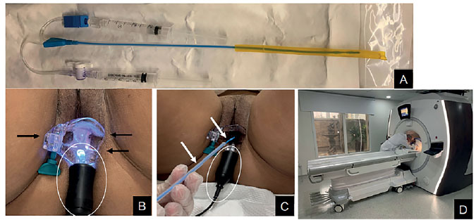
Figure 1. MR-HSG equipment
A: MR-HSG equipment.
Black circle: end of the catheter with the balloon that will be placed in the uterine cervix.
Blue circle: syringe used to inflate the catheter.
Yellow: syringe used to inject the contrast medium (gadolinium + saline).
B and C illustrate items used: black arrows indicate the plastic speculum (MR compatible). The white circle shows the light connector for better view of the external orifice of the uterine cervix. White arrows show the HSG equipment as it is introduced in the vagina.
D: patient positioned at the MR table as the radiologist administers the contrast medium.