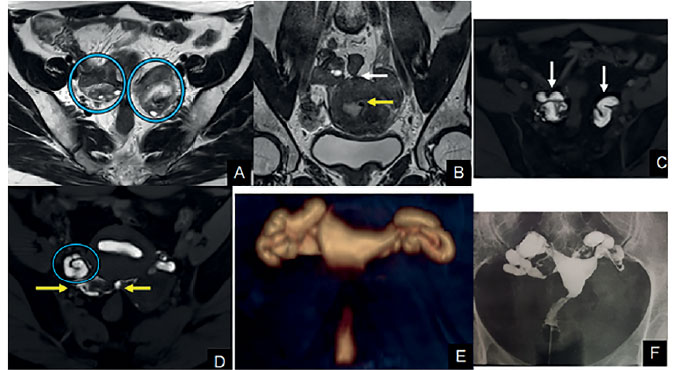
Figure 2. Exam example
A: MR T2WI in the axial plane. The circles show ovaries close to the midline in the posterior pelvic pouch and adhered to the retrouterine endometriotic tissue. B: T2WI in the coronal plane. The yellow arrow shows a small submucosal fibroid measuring 3 mm; the white arrow indicates retrouterine endometriosis with adhesions involving both the ovaries and the rectum. C: Dynamic MR-HSG sequence in the axial plane. White arrows show bilateral hydrosalpinx with mucosal thickening. D: The circle shows right tube accumulated in the posterior pelvic pouch. E: MR-HSG volume rendering showing bilateral hydrosalpinx. F: Conventional HSG with bilateral hydrosalpinx.