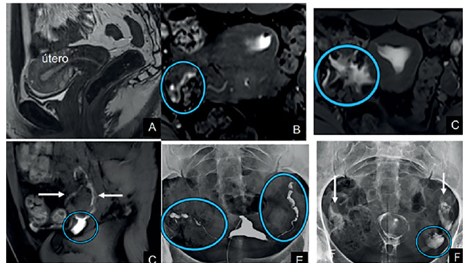
Figure 3. Exam example A: T2WI in the axial plane showing no abnormalities. B: Dynamic MR-HSG in axial plane. The circle shows part of the right tube, normal appearing. C: Dynamic MR-HSG axial plane. The arrow shows the uterine cavity filled with contrast and the circle indicates free tubal spillage to the peritoneal cavity. D: Dynamic MR-HSG in the sagittal plane. The arrow demonstrates the left ovary with a small amount of adjacent fluid. The circle shows a small amount of fluid in the left ovarian fossa. E: Conventional HSG showing normal tubes (circles). F: Conventional HSG, late phase (Cotté). The arrows demonstrate normal contrast dispersion in the peritoneal cavity. The circle shows periovarian fluid.