JBRA Assist. Reprod. 2020;24(4):518-524
ORAL PRESENTATIONS
doi: 10.5935/1518-0557.20200072
Abstracts of the 24th Annual Congress of the SBRA, 2020 (Virtual Meeting)
O-01. Trisomy causes accelerated embryonic development. Is that an indication for preimplantation genetic test for aneuploidy in in vitro fertilized embryos?
1Centro de Reprodução Humana Monteleone, São Paulo. Brazil2Disciplina de Ginecologia - Departamento de Obstetrícia e Ginecologia. Faculdade de Medicina da Universidade de São Paulo (FMUSP), São Paulo. Brasil
3Departamento de Ginecologia. Escola Paulista de Medicina da Universidade Federal de São Paulo (EPM-UNIFESP), São Paulo. Brazil
ABSTRACT
Objective: Human embryos are affected by a high rate of aneuploidy, around 35% in natural conceptions and this prevalence is expected to be higher in pre-implantation embryos derived from in vitro fertilization (IVF). One of the most common alteration is the trisomy and preimplantation genetic testing for aneuploidies (PGT-A) is currently used in IVF to minimize the chances of transferring genetically abnormal embryos. The introduction of time-lapse monitoring (TLM) allowed additional noninvasive criteria to best embryo selection. Markers of embryonic kinetic from the first-cleavage onwards has been studied and applied in algorithms for best embryo choice. The aim of this study was to analyze the association of trisomy with the embryo morphokinetics in embryos derived from IVF and analyzed by PGT-A.
Methods: This retrospective study included 509 embryos from 109 patients undergoing IVF between April/2018-September/2019. Mature oocytes were fertilized by ICSI and cultured in a TLM system (Embryoscope®). Blastocysts were biopsied for PGT-A and analyzed at Igenomix Brasil through Next Generation Sequencing (NGS). Twenty-seven embryos presenting trisomy of chromosomes 13, 15, 16, 18, 21 and 22 were selected and a matched analysis was performed, since a euploid embryo from the same patient was selected as a control for each trisomic embryo.
Results: The women age was 38.0±3.0 years and euploid blastocyst rate was 36.3%. Two-way repeated measured ANOVA was performed based on times for achieving pronucleous-fade, two-cells, three-cells, four-cells, five-cells, eight-cells, nine-cells and blastocyst. Embryo morphokinetics significantly differed between euploid and trisomic embryos (p=0.052). The pairwise comparisons demonstrated significant difference on times between euploid and trisomic embryos from eight cells until blastocyst stage. The mean difference time between euploid and trisomic embryos to get the eight-cells stage was 5.1 hours (61.0±10.8x55.9±7.1, p=0.033) and to attain blastocyst stage was 6.2 hours (112.3±11.7x106.1±10.4, p=0.013).
Conclusion: In this study, trisomic embryos showed a faster development than their paired euploid ones. Despite of this study have included a small number of embryos, the inclusion criteria was for each trisomic embryo there is an euploid embryo of the same patient, which exclude a number of confounders. The literature describes the association of accelerated embryonic development with lower success potential; however, the reason for that relationship is not clear. Our findings allow us to hypothesize that the decreased implantation potential of faster embryos could be due to chromosomal alteration, specifically trisomy, and to suggest that accelerated embryonic development may be an indication for genetic evaluation. These findings must be confirmed in a higher number of embryos and further studies are being conducted to investigate the association of genetic embryo alterations and embryo development.
O-02. Motherhood plan: has it changed in face of the COVID-19 pandemics?
1Fertility Medical Group - São Paulo/SP2Instituto Sapientiae - Centro de Estudos e Pesquisa em Reprodução Assistida- São Paulo/SP
ABSTRACT
Objective: The novel coronavirus (Covid-19) outbreak led to a public health emergency of international concern, putting health organizations on alert. World authorities implemented suppression plans to control community spread, including restrictions to non-urgent medical care. Assisted reproduction centers had to adapt to these restrictions. The infertility diagnosis and reproductive treatments possess an inherent psychological burden. This associated with the uncertainty of the consequences of the passage of time in the prognosis of treatments may impact on patient’s psychological health. The goal for the present study was to investigate whether women seeking fertility care have different perception concerning the impact of Covid-19 on the motherhood plan than a target population?
Methods: From 22/April/2020 to 25/may/2020, a survey through an online-platform was conducted. Participants were randomized by age, in a 1:4 ratio, into one of the two groups: ART-GROUP (n=368), including patients seeking for fertility treatment, but still didn’t start their cycles or INTERESTED-GROUP (n=92), including participants interested in the subject, who accessed the website of a university-affiliated IVF-center. Participants in the ART-GROUP were invited via e-mail, with a cover-letter outlining the survey and a link to access it. Participants in the INTERESTED-GROUP accessed the questionnaire via website. Information on demographic data and their perceptions in face of the COVID-19 pandemics and the motherhood plan was collected. Women were asked: (i) How do you see the possibility of becoming pregnant after the beginning of the COVID-19 pandemic? (ii) How long do you think that suppression strategies will last? and (iii) Did you postpone your plans to become pregnant? If yes, why? Subjects would choose more than one reason to postpone the plan to become pregnant.
Results: Most patients seeking for fertility treatment were married or in a common-law relationship (73.1% vs. 48.9%, p<0.001, for ART-GROUP and INTERESTED-GROUP, respectively). When asked about the possibility of becoming pregnant, after the beginning of the pandemic, 40.7% of the ART-GROUP stated to believe the pandemic could affect their plans, while only 31.5% of the INTERESTED-GROUP stated the same (p=0.067). Concerning the duration of the suppression strategies, patients in ART-GROUP were more optimistic: 45.4% of ART-GROUP and 80.4% of INTERESTED-GROUP stated to believe the suppression strategies will last at until August or more (p<0,001). The plan to become pregnant was postponed by 59.8% of the ART-GROUP and by 43.4% of the INTERESTED-GROUP (p<0.001). The main reasons that led people to this decision included fear of getting sick (59.2% vs. 66.3%, p=0.435, for ART-GROUP and INTERESTED-GROUP, respectively), economic reasons (42.4% vs. 27.1%, p=0.086 for ART-GROUP and INTERESTED-GROUP, respectively) and a pessimist view of the future (13.8% vs. 23.9%, p=0.073 for ART-GROUP and INTERESTED-GROUP, respectively).
Conclusion: Couples seeking fertility care have a different perception regarding the impact of COVID-19 on the parenthood plan than a target population. Besides the fear of becoming sick and pessimist view of the future, economic burdens seen to be the main reason for the delay in the parenthood plain, which may be due to the fear of future economic instabilities and the fact that, in Brazil, ART do not qualify for reimbursement.
O-03. Can we predict aneuploidy with morphokinetics parameters?
1Huntington Medicina Reprodutiva, Embryology, São Paulo, Brazil.2Huntington Medicina Reprodutiva/Federal University of São Paulo, Clinical Director /Associate Professor, São Paulo, Brazil.
3Huntington Medicina Reprodutiva, Research & Development, São Paulo, Brazil.
ABSTRACT
Objective: The success of an IVF treatment lays on selecting for uterine transfer the embryo with the best potential to develop a healthy baby. Currently, embryo ploidy status is only known by analyzing biopsied cells, an invasive and expensive procedure. Concerning non-invasive analysis, aneuploidy may affect embryo morphokinetics, but among the several studies reported, no morphokinetic parameter has been consistently identified as predictive of embryo ploidy status. The objective of this study is to investigate if there are differences in morphokinetic parameters between euploid and aneuploid blastocysts.
Methods: This was a retrospective cohort study that evaluated the morphokinetic parameters of 593 biopsied blastocysts from patients undergoing IVF with clinical indication for Preimplantation Genetic Test for Aneuploidy (PGT-A) and analyzed by Next Generation Screening platform (NGS), from autologous cycles cultured between December 2017 and December 2019 in a private IVF center. Maternal age included was limited between 37 and 42 years old (n= 191 patients). All oocytes were fertilized by Intra Cytoplasmatic Sperm Injection (ICSI) and cultured in time-lapse system incubator (Embryoscope Plus, Vitrolife). Embryos were analyzed according to the following morphokinetic parameters, annotated in hours (h): time of pronucleous fading (tPNf), time to 2-cell(t2), time to 3-cell (t3), time to 4-cell (t4), time to 5-cell (t5), time to 8-cell (t8) and time to blastulation (tB). Embryos were graded according to Gardner’s morphology parameters. Statistical significances were calculated using chi square or t-test as appropriated.
Results: Five-hundred ninety-three blastocysts were analyzed. From those, 227 were euploid (38%) and 366 aneuploid (62%). Maternal age was similar between euploid and aneuploid embryos (39.34±1.46 versus 39.57±1.45, p=0.0704). Morphokinetics parameters were faster in euploid embryos in two time-points: tPNf and tB (tPNf: 23.55h±2.94h versus 24.07h±3.27h, p=0.0406; tB: 108.21h±9.94h versus 111.21h±11.51h, p=0.0003). Regarding other parameters analyzed, all were similar between euploid and aneuploid embryos (t2: 26.44h±3.26h versus 26.91h±3.15h, p=0.0533; t3: 37.13h±3.97h versus 37.41h±3.97h, p=0.1375; t4: 38.42h±4.48h versus 38.74h±4.30h, p=0.2036; t5: 49.21h±6.47h versus 49.74h±6.86h, p=0.2805; t8: 58.48h±9.01h versus 59.58h±9.23h, p=0.117). Morphology grades between euploid and aneuploid embryos were different between good quality embryos (grades A and B - 82% versus 63%) and poor quality embryos (at least one grade C - 18% versus 37%, p<0.0001). These results are described in table 1.
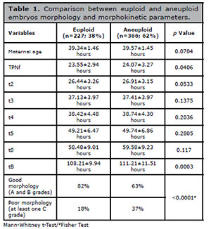
Table 1. Comparison between euploid and aneuploid
embryos morphology and morphokinetic parameters.
Conclusions: Our data identified that euploid blastocysts achieved time of pronucleous fading (tPNf) and time to blastulation (tB) earlier than aneuploidy blastocysts, and this endpoint can be useful in recommending and indicating PGT-A for patients. As non-invasive techniques to access embryo biological potential are emerging in IVF laboratories, associating morphokinetic parameters and ploidy status is another potential tool to improve embryo selection, with the perspective of removing embryo biopsy in the future.
O-04. Early and late paternal contribution to cell division of embryos in a time-lapse incubation system
1Fertility Medical Group - São Paulo/SP2Instituto Sapientiae - Centro de Estudos e Pesquisa em Reprodução Assistida - São Paulo/SP
ABSTRACT
Objective: In assisted reproductive treatment (ART), time-lapse imaging (TLI) offers a non-invasive observation of morphokinetics parameters under stable culture condition, which have improved embryo development and allowed the development of numerous algorithms for the selection of embryos for transfer and prediction of embryo implantation. Previous studies have demonstrated that embryo development is affected not only by intrinsic embryonic quality, but also by parental factors, such as maternal and paternal ages, ovarian stimulation, semen parameters, embryo culture media and so on. However, studies on the impact of paternal factors on morphokinetics events are still scarce. The identification of morphokinetics events affected by paternal factors may contribute to a better understanding of morphologic mechanisms of fertilization and behavior of human embryos. The aim of this study was to investigate the impact of paternal age and semen quality on embryo morphokinetics events in a TLI incubator.
Methods: This cross-section study was performed in a private university-affiliated IVF center, between March and November/2019. Kinetic data were analyzed in 1220 embryos individually cultured in a TLI incubator until day five of development, originating from 139 patients undergoing ICSI. Timing of specific events from the point of insemination was determined using time-lapse imaging. The incubator high-definition camera was set up to record embryos’ images, in eleven focal planes, every 10 minutes. Recorded kinetic markers were: timing to pronuclei appearance (tPNa) and fading (tPNf), timing to two (t2), three (t3), four (t4), five (t5), six (t6), seven (t7), and eight cells (t8), and timing to blastulation (tB). Durations of the second (t3-t2) and third (t5-t3) cell cycles (cc2 and cc3, respectively) and timing to complete synchronous divisions s1 (t2-tPNf), s2 (t4-t3), and s3 (t8-t5) were also calculated. Abnormal cleavage patterns, such as reverse cleavage and direct uneven cleavage, and the presence of multinucleation were also recorded. Multivariate linear regression analysis, adjusted for maternal age, was used to evaluate the influence of male age, ejaculatory abstinence length and semen parameters on embryo morphokinetics events and clinical outcomes.
Results: The paternal age was directly correlated with longer t2 (B: 0.043, CI: 0.006 - 0.081, p=0.043), t3 (B: 0.056, CI: 0.001 - 0.110, p=0.044), t4 (B: 0.066, CI: 0.014 - 0.118, p=0.012), t6 (B: 0.080, CI: 0.002 - 0.163, p=0.046), tB (B: 0.195, CI: 0.003 - 0.387, p=0.046), and was also associated higher incidence of multinucleation (Exp(B): 1.027, CI: 1.043 - 1.203, p=0.004) and abnormal cleavage patterns (Exp(B): 1.116, CI: 1.034 - 1.205, p=0.005), while the implantation rate (B: -1.933, CI: -2.426 - -1.441, p<0.001) and the odds of pregnancy (Exp(B): 0.899, CI: 0.860 - 0.940, p<0.001) were negatively affected by paternal age. The length of EA was inversely correlated with implantation rate (B: -0.014, CI: -0.026 - -0.002, p=0.027). The sperm count was correlated with shorter s3 (B: -0.005, CI: -0.010 - -0.001, p=0.026). The progressive sperm motility was correlated with shorter t4 (B: -0.016, CI: -0.032 - -0.001, p=0.037), t6 (B: -0.028, CI: -0.052 - -0.003, p=0.027), t7 (B: -0.044, CI: -0.073 - -0.015, p=0.003), t8 (B: -0.054, CI: -0.087 - -0.021, p=0.002), s3 (B: -0.033, CI: -0.060 - -0.006, p=0.019), and tB (B: -0.050, CI: -0.089 - -0.012, p=0.010). The TMSC was correlated with shorter t8 (B: - 0.009, CI: -0.017 - 0.001, p=0.021), S3 (B: - 0.009, CI: -0.016 - -0.003, p=0.005), and tB (B: - 0.011, CI: -0.020 - -0.002, p=0.019).
Conclusion: Increasing paternal age and poor seminal quality correlate with delayed cell cleavage and blastulation. These findings add to the general knowledge of the impact of paternal factors on embryo development, and highlight the importance of paternal contribution for the ART success. Results found here may be helpful in recommending shorter EA periods, and provide support for day 5 embryo transfer in couples with advanced male age and or poor semen quality.
O-05. Oocyte ability to repair sperm DNA fragmentation: The effect of maternal age on ICSI outcomes
1Fertility Medical Group - São Paulo/SP2Instituto Sapientiae - Centro de Estudos e Pesquisa em Reprodução Assistida - São Paulo/SP
ABSTRACT
Objective: Sperm DNA integrity is crucial for the adequate transmission of paternal genetic information and has been recognized as a biomarker of male infertility. Previous studies have suggested a negative association between sperm DNA fragmentation (SDF) index and pregnancy after ICSI, while others, despite having failed to find such association, have suggested an association with pregnancy loss. Spermatozoa have no mechanism to repair SDF, therefore, the DNA-repairing activity depends on the oocyte machinery once fertilization takes place. The SDF-repairing oocyte’s ability depends not only on the level of SDF, but also on the quality of the oocyte. Increasing maternal age has a well-established negative effect on oocyte quality that reflects in fertility and reproductive success. Therefore, the goal for the present study was to evaluate the impact of SDF on clinical outcomes of assisted reproductive technology in women in different age groups.
Methods: This study included data from 540 ICSI cycles, performed from June/2017 to December/2019, in a private university-affiliated IVF center. Cycles were split into three groups according to the maternal age: ≤36 years old (n=285), 37-40 years old (n=147), and >40 years old (n=108). Semen samples were evaluated for SDF using the Sperm Chromatin Dispersion method and for each age group, the cycles were divided again according to SDF index: low fragmentation index (≤30% SDF) and high fragmentation index (>30% SDF). Clinical outcomes were compared between the groups using generalized linear models followed by Bonferroni post hoc test, with adjustment for potential confounders.
Results: For younger patients (≤36 years old) and those between 37 and 40 years old, no significant differences were noted in pregnancy (≤36 years old: 40.0% vs. 39.1%, p=0.840 and 37-40 years old: 27.7% vs. 28.6%, p=0.781), implantation (≤36 years old: 42.3% vs. 41.5%, p=0.880 and 37-40 years old: 28.9% vs. 30.6%, p=0.757), or miscarriage rates (≤36 years old: 9.3% vs. 11.1%, p=0.665 and 37-40 years old: 31.2% vs. 22.2%, p=0.875), for cycles with ≤30% SDF or >30% SDF respectively. However, when maternal age was >40 years old, significant lower pregnancy (20.0% vs. 7.7%, p=0.040) and implantation rates (19.7% vs. 11.9%, p=0.040), and increased miscarriage rate (12.5% vs. 100.0%, p<0.001) were observed for cycles with ≤30% SDF or >30% SDF, respectively.
Conclusion: High SDF index leads to lower implantation and pregnancy rates and higher miscarriage rates in ICSI cycles of women with advanced maternal age. The same is not observed when maternal age is < 40 years old. This evidence indicates that, when the oocyte is injected with a DNA-damaged spermatozoon, the maternal age may affect the oocyte DNA-repairing ability, leading to the development of an embryo with both lower implantation and higher miscarriage potentials.
O-06. Zika virus infectivity in human ovarian granulosa cells
1Institute of Biophysics Carlos Chagas Filho, Universidade Federal do Rio de Janeiro (UFRJ), Rio de Janeiro, RJ, Brazil.2Fertipraxis Centro de Reprodução Humana, Centro Médico Barrahopping - Rio de Janeiro, RJ, Brazil.
3Institute of Microbiology Paulo de Góes, Universidade Federal do Rio de Janeiro (UFRJ), subsolo - Rio de Janeiro, RJ, Brazil.
ABSTRACT
Introduction: Anvisa regulates the preservation and acquisition of gametes in assisted human reproduction clinics by the resolution number 23 (RDC23, May 27th, 2011). All patients must be tested for a range of pathogens. After the Zika virus (ZIKV) outbreak in 2016, Anvisa updated this resolution by adding ZIKV tests (RDC72; March 30th, 2016). These tests generate more expenses and concerns for patients and doctors at the clinics. Human granulosa cells (HGC) represent an important selection barrier for substances and pathogens for the oocyte. However, there is little evidence in the literature about the infectivity of HGC by ZIKV. A cell can be susceptible to a virus if it can penetrate the cell and can be permissive if the cell replicates the viral genome and assemble infectious viral particles. The present study aims to investigate the infectivity of HGC by ZIKV.
Methods: For that purpose, we used two HGC models: the COV434 strain and HGC isolated (HGCi) from patients submitted to assisted reproduction techniques in a single center. The CGHi were obtained from the follicular aspirate of patients with at least 10 follicles with a diameter ≥14mm. The cell clusters were selected manually with a micropipette and tranfer to PBS buffer supplemented with synthetic serum. The samples were transported to the university laboratory for further isolation. In the laboratory the sample was treated with hyaluronic acid to break up cell clusters and with ACK lysing buffer for red blood cells. After successive centrifugations, the cells were plated and cultured in supplemented medium. In order to assess the infectivity of cells by ZIKV, viral tropism was analyzed according to the concepts of susceptibility and permissiveness. In the infection tests, two viral strains were used: the African (ZIKV/MR-766) and the Brazilian (ZIKV/PE-243). Also, we evaluated three different multiplicities of infection (MOI): 1, 5 and 10, which means number of viral particles per cell. Supernatants from cultures treated with ZIKV were collected on day 0 (input) and day 3 for titration by plaque and qPCR assay. Cell viability was analyzed by the XTT assay kit (Sigma Aldrich) and by excluding trypan blue. The XTT colorimetric assay system is used to measure cellular metabolic activity as an indicator of cell viability.
Results: We observed the presence of viral RNA in the cytoplasm of COV434 as well as an increase in viral load in the supernatant after three days of infection with both strains. However, the cytopathic effect was only observed in MOI 10 by ZIKV/MR-766 (60% viability, p<0.05). Taking together, our data demonstrated that COV434 is both susceptible and permissive to ZIKV. Additionally, the presence of viral RNA was positive in the HGCi cytoplasm. However, it was not possible to observe the formation of new viral particles in the supernatant of these cells after three days of infection. Similar to COV434, the viability of HGCi was compromised only with ZIKV/MR-766 in MOI 10. Thus, we could demonstrate that HGCi are permissive to ZIKV. We also analyzed the presence of membrane receptors discussed in the literature as possible routes of entry for ZIKV, which were present in both cell models.
Conclusion: In the present study, we concluded that both COV434 and HGCi have key receptors for ZIKV entry and are susceptible to it. However, the permissiveness was observed only in the lineage model. A limitation of the study failed to prove whether the HGCi model is permissive to the virus, since we are not able to observe with certainty the presence or absence of ZIKV in cells. Further studies are needed to confirm that ZIKV can not replicate in HGCi.
O-07. Validation process of free-DNA medium collection for noninvasive preimplantation genetics test for aneuploidy (niPGT-A)
1Paulista Center for Diagnosis-Research and Training, Ribeirao Preto, Brazil.2Centre for Human Reproduction Prof. Franco Jr, Ribeirão Preto, Brazil.
3São Jose do Rio Preto School of Medicine FAMERP, São Jose do Rio Preto, Brazil.
4Ferticlin Human Fertility Clinic, Research, São Paulo, Brazil.
5Materbaby, Maringa, Brazil.
6Santista Nucleus of Human Reproduction, Santos,Brazil.
7Feliccita Fertility Institute, Curitiba, Brazil.
8Genesis Human Reproduction Assistance Center, Brasilia, Brazil.
9Fertility Medical Group, Research, São Paulo, Brazil.
10Art Fertil Human Reproduction Clinic, Recife,Brazil.
11Alpha Project - Alliance of Assisted Fertilization Laboratories, São Paulo, Brazil.
12CENAFERT, Salvador, Brazil.
ABSTRACT
Objective: To validate the collection processes of free-DNA, released by the embryo to the culture media during their development, for future niPGT-A evaluation.
Methods: This is a multicenter study performed by a total of 11 Brazilian assisted reproduction centers (Bra-ARC). The validation process was performed into two steps; first step (frozen embryos): collection of the medium in which frozen/thawed embryos were cultured and second step (fresh embryos): collection of the medium in which fresh embryos were cultured. For the first step: each center thawed at least 3 blastocysts (Day5/6), mean of 5.4±3.1, previously biopsied for invasive PGT-A (iPGT-A) and diagnosed as genetically abnormal with consent for disposal. A total of 44 frozen-thawed biopsied blastocyst embryos were cultured in individual wells of 15µl of culture medium in GPS dishware (MGPS010 Global) until they reached their expansion (4-24 hours). Expanded blastocysts were individually transferred to PCR tubes and their corresponding free-DNA spent medium were collected for niPGT-A evaluation. In this step, it was possible a comparison between the genetic diagnosis of the whole embryo (gold stander), with the free-DNA released by the embryo into the culture medium during their development and the diagnosis of previous trophectoderm biopsy results. For the second step: each center cultured at least 3 fresh cleaved (D3) embryos, mean of 5.1±2.9. A total of 40 embryos were transferred into individual well of 20 µl of culture medium in GPS dishware and were cultured from day 3 to day 5/6. Expanded blastocysts (grade 4) were vitrified and their respective spent culture medium were collected and transferred to PCR tubes for niPGT-A evaluation.
Results: A total of 84 samples of all Bra-ARC (from both first and second steps) were used for niPGT-A. From 74 sequenced samples, 26 embryos were euploids (35.1%), 38 embryos presented aneuploidy (51.4%) and 10 of them (13.5%) were mosaics. The sex rate was 55.4% for XY and 44.6% for XX. Ten samples (11.9%) failure sequencing for insufficient or poor DNA quality samples (table 1). The mean of free-DNA concentration into the collected spent media was 29.8 ng/µl and 19.2ng/µl for the first and second step, respectively. From the first step (Table 1): From 46 Free-DNA samples and whole embryos, 44 (95.6%), were successfully amplified. Comparing the results of niPGT-A and whole embryos sequencing, the predictive value of the normal test was 100% (PVNT) and the false abnormal test (FAT) was 8.3%. On the other hand, when we compared the whole embryos and iPGT-A, false abnormal test was 25%, evidencing that the biopsy procedure increased 3 times the FAT rates compared with niPGT-A (no biopsy procedure). From the second step: 73% of the medium was collected on D5 and only 27% of them were collected on D6, shortly thereafter the blastocyst reached grade 4. At the beginning of the validation process, 80% of all free-DNA spent medium evaluated presented enough free-DNA concentration to performed niPGT-A. Therefore, it increased for 96.9% after Bra-ARC validation process (data sent for publication). The mean DNA concentration collected by each center varied from 2.2 to 59.4 ng/µl, depending of blastocyst grade and day of collection. (D5/D6). From 32 spent free-DNA samples assessed, 40.6% (13/32) of them presented euploidy, 46.8% (15/32) aneuploidy, 12.5% (4/32) mosaicism.
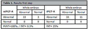
Table 1. Results first step
Conclusion: The validation process showed to be a powerful procedure for future niPGT-A evaluation. All of the Bra-ARC has shown effective for the collection process of free-DNA for both protocols (fresh or frozen embryos culture), after protocol adjusting. The niPGT-A showed 3 times less false abnormal rates compared to iPGT-A.
O-08. Surrogacy: the main indications and couple’s biggest challenges, an 18-year psychology experience in a reproductive medicine clinic in Brazil.
1Huntington/Pró-criar Medicina ReprodutivaABSTRACT
Objective:
Demonstrate through numbers the main indications of surrogacy, the difficulties encountered by the couple and how the psychologist’s action helps to overcome these obstacles. Methods: Retrospective analysis of the psychological follow-up of patients who sought an in vitro fertilization treatment with temporary uterus donation highlighting aspects related to medical indication, psychological assessment and ethics discussion.
Results: A total of 77 patients had the indication of surrogate between April 2003 and April 2020 at Pró-Criar reproductive medicine. Of these, 30 (39%) actually underwent treatment, which corresponds to a conversion rate of 39%. The average age of people who underwent treatment was 35 years. The main medical indications for surrogacy were following hysterectomized women for benign diseases in 23 cases (29,9%);serious medical conditions in 17 (22%);12 homosexual patients (15,6%);congenital absence of uterus in 11 (14,3%); cancer surgery in 6 patients (7,8%);recurrent miscarriage in 6 cases (7,8%) and repeated IVF failures in 2 cases (2,6%). Of the 30 couples undergoing treatment, 13 (43,3%) were able to conceive and have a live birth;11 (36,7%) were negative results; 4 (13,3%) progressed to abortion and 2 (6,7%) had a canceled cycle. We had incomplete data from 9 (11,7%) patients and another 34 (44,1%) did not undergo treatments after the indication. The main reasons for not undergoing treatments were:18 (53,0%) had a first medical and psychological evaluation, but chose not to undergo the treatment; 9 (26,5%) was considered unfit by the doctor; 4 (11,8%) were considered psychologically unfit; 1 (2,9%) gave up the treatment, due to the couple broke-up; 1 (2,9%) would have to use donated eggs with surrogacy and this was not authorized in CFM Resolution number 2.013/2013 and 1 (2,9%) was not authorized by the CRM / MG, as the patient was a single man and there was no inclusion of single people in chapter VII of Substitute gestation at CFM Resolution no.2.121 / 2015. Four patients (5,2%) were underwent oocyte cryopreservation for future treatment. The action of psychologists was fundamental, especially in cases of clarifying the doubts of surrogacy, demonstrating the natural weaknesses of a pregnancy as in the case of abortions, in the careful assessment of the couple’s psychological profile and the consequences for them in a pregnancy by surrogate, in the understanding of medical refusal and class advice through their codes of ethics and mainly be available throughout the treatment process for possible doubts that arise. In Brazil, the CFM, in its resolution no. 2.168/2017 determines that the patient’s medical record must contain a medical report, with a psychological profile, attesting the clinical and emotional adequacy of all those involved, and this has been the practice at Pro-Criar reproductive medicine since the first case of surrogacy in 2003.
Conclusion: The surrogacy is an alternative treatment that gives patients, unable to conceive, can have a biological child, provided that everyone involved in the process is well evaluated from a medical, psychological and ethical point of view.
O-09. Three-dimensional (3D) in vitro culture using magnetic levitation system of bovine ovary fragments maintain tissue viability and balanced oxidative status
1Gynecology and Obstetrics Program at Ribeirão Preto Medical School, University of São Paulo.2Veterinary Sciences Program at the University of Uberlândia.
3Sapientiae Institute - Center for Study and Research
4Institute of Biomedical Sciences, University of Uberlândia.
5Centro Universitário do Triângulo - UNITRI, Uberlândia.
6Veterinary, no institucional affiliation.
7Ribeirão Preto Medical School, University of São Paulo.
ABSTRACT
Objective: The aim of this study was to evaluate the viability, mitochondrial activity and oxidation of bovine ovarian tissue cultured in three different 3D systems: 1. magnetic levitation (LM), 2. alginate matrix (AG) or 3. fibrin-alginate matrix (FA).
Methods: Ten fragments were included in each in vitro culture group, and 4 fragments were collected as fresh control group, thus a total of 34 fragments. These ovarian pieces were cultured in LM culture (nanoparticle of gold and iron oxide was cross-linked by poly-L-lysine, which is a cell-adhesive peptide sequence) (400 µL/mL) or AG 1% or FA 1%. In each group the fragments were subdivided for analysis in day 1 (D1-N=5) and in day 7 (D7-N=5). Confocal analyzes was performed immediately after sample collection (D0- pre-culture), 24 hours (D1) and seven days (D7) after cultivation. The fragments were evaluated for signs of cell degeneration (propidium iodide method), mitochondrial activity (MitoTracker Orange CMTMRos technique) and intercellular levels of production of reactive oxygen species (ROS) (DCF method). All markers were analyzed by confocal microscopy and measured as the numbers of pixels obtained, controlling for background staining. After averaging the pixels of the images obtained by the confocal microscope, each fragment was analyzed at 5 different points and at each point, the 3 probes (propidium iodide, MitoTracker Orange CMTMRos and DCF) were evaluated. Two analyzes were performed for each variable: 1) One-way ANOVA for comparisons in relation to the Control group; 2) Two-way ANOVA to assess the effects of treatments (3D culture by magnetic levitation, alginate and alginate with fibrin) and days (1 and 7).
Results: When assessing tissue viability (degeneration), we observed that tissues cultured in LM system maintained viability similar to controle group (p>0.05), while AF and AG were slightly higher than control (p<0.05) (Table 1); however after 7 days of culture LM and AF had similar viability, both comparable to control (p>0,05) and AG had higher degeneration rates (p<0.05). When analyzing the mitochondrial activity, the LM system had similar results to the control group, which was lower than the AG group (p<0.05); the AF group had intermediate mitochondrial activity not differing from LM, control or AG groups (p>0.05). This pattern of mitochondrial activity did not differ between days 1 and 7 in any of the 3 culture groups. Finally, when analyzing the production of ROS (redox status) we observed that the control samples presented with levels of ROS production (DCFH), probably due to all tissue transportation and processing. When these tissues were cultured there was a reduction in ROS production in groups LM and AG in D1 (p<0.05 in comparison to control), indicating some degree of recovery in its oxidation balance, but the AF group maintained high levels of ROS (p>0.05, when compared to control). AG group did reduce its ROS production on D7, with leves similar to LM system (both lower than control, p<0.05), however AF group presented with an increase in ROS production which was similar to the control group (p>0.05).
Conclusions: The LM system presented the best results for in situ cultivation with viability similar to fresh control tissue, low mitochondrial activity similar to control, which was probably enough to maintain intracellular oxidative balance as they presented with the lowest levels of ROS emission among all groups, even when compared to the control group. AF group also had good results in terms of tissue viability and mitochondrial activity, however it was not capable of maintaining proper redox status when ROS production was analyzed.
O-10. Granulosa cell signaling based bioassay in vitro for personalized stimulation in ART
1University of Modena and Reggio Emilia, Unit of Endocrinology-Dept. Biomedical-Metabolic and Neural Sciences, Modena, Italy2Arcispedale Santa Maria Nuova, Department of Obstetrics and Gynaecology-Fertility center, Reggio Emilia, Italy
3Merck KGaA, Merck Serono S.p.A - Rome- Italy, Darmstadt, Germany
ABSTRACT
Study question: Is the individual response to gonadotropins predictable?
Summary answer: Granulosa cells (hGLC) from normo-responder women display higher cAMP and progesterone production in vitro, as well as estradiol levels in vivo, than sub-responders.
What is known already: The various stimulation protocols do not provide the best stimulation setting and some patients may be subjected to inadequate stimulation resulting in clinical treatment failure. It is expected that hGLC discarded samples donated by normo-responder and sub-responder women undergoing assisted reproduction (ART) feature specific follicle-stimulating hormone (FSH) receptor (FSHR) expression and response to gonadotropins in vitro, suggesting they may be used for predicting the response in vivo of normo- versus sub-responder women.
Study design, size, duration: hGLC samples from anonymized donor women undergoing oocyte retrieval for ART are collected during a 30-months period (2017-2019). Cells are purified, cultured, genotyped and treated by gonadotropins in vitro and intracellular signaling endpoints are collected. Data obtained by cells from 105 normo-responder women are compared to those from 105 sub-responders, which are identified as patients providing ≤4 oocyte retrieved. Experiments are performed under the local Ethics Committee permission (n◦2018/0080377,16/07/2018) and donor written consent.
Participants/materials, setting, methods: hGLC donor women undergone ART without endocrine abnormalities and infectious diseases. hGLC samples are genotyped for FSHR polymorphisms known to modulate the response to FSH (rs6166, rs6165, rs1394205) and stimulated by increasing doses (nM range) of recombinant FSH (Gonal-F, Merck KGaA, Darmstadt, Germany). cAMP and progesterone are measured by immunoassays and dose-response curves performed. Results from samples will be used as reference Control Group and/or compare different sample populations, together with clinical data.
Main results and the role of chance: To date, 76 samples resulted to be eligible for in vitro stimulation. 3-h cAMP data from normo- versus sub-responder women resulted in different potency, as demonstrated by 50% effective concentrations (EC50) in vitro (FSH EC50 normo-responders=5.8Å}4.9 nM; EC50 sub-responders=2.4Å}9.8nM; p<0.05; t-test), and different plateau levels, indicating phenotype-specific e cacy (cAMP plateau normo-responders=37.34Å}5.3 pmol/ml; cAMP plateau sub-responders=15.69Å}4.2 pmol/ml). 24-h basal progesterone production of cells from normo-responder women is about 1.3-fold higher than that of cells from sub-responders. Most importantly, both the groups of data confirmed that FSH elicit phenotype-specific response, depending on the normo- or sub-responder status. Indeed, FSH EC50, and progesterone basal and plateau levels are higher in cells from normo-responders versus sub-responders (p<0.05; t-test). Distribution of FSHR genotypes was evaluated by contingency analysis. No different allele frequencies and FSHR expression levels between normo- versus sub-responders were found (Chi square and t-test; p>0.05; n=55), demonstrating that each the two groups are genetically homogeneous. Estradiol levels are higher in normo- than sub-responders (2026Å}903.5 versus 1375Å}898.9pg/ml; t-test; p>0.05; n=49).We demonstrate that normo-responders display higher sensitiveness to FSH than sub-responders, revealing association between intracellular signaling, estradiol production and follicle growth.
Limitations, reasons for caution: To date, patient recruitment is still ongoing and about one hundred individuals should be required to achieve proper statistical power.
Wider implications of the findings: This study may predict the individual response to personalize ART protocol, in case of stimulation cycle planning after previous failures. Given the different in-vitro response between cells from normo- vs sub-responders, we expect that this in-vitro assay could lead to future clinical trials that may confirm in vitro results.
Trial registration number: not applicable
Abstract presented (Oral Presentation O-282) at the 35th Annual Meeting of the ESHRE, Vienna, Austria 24 to 26 June 2019. Published by permission of Oxford University Press on behalf of ESHRE Oxford University Press License Number: 4851800745400