JBRA Assist. Reprod. 2020;24(4):391-393
EDITORIAL
doi: 10.5935/1518-0557.20200075
Noninvasive preimplantation genetic testing for aneuploidies (niPGT-A) and the principle of primum non nocere
1Center for Human Reproduction Prof. Franco Jr., Ribeirão Preto, SP, Brazil
ABSTRACT
Perhaps with the intention of obtaining larger amounts
of free-DNA, some groups are routinely postponing and
establishing free-DNA collection in culture medium for
Noninvasive preimplantation genetic testing for aneuploidies
(niPGT-A) to day 6 for all blastocysts. A meta-analysis
served as the basis for such decision, since statistically
similar live birth rates were observed when the transfers of
euploid blastocysts were performed on day 5 versus day 6
However, the euploidy analysis was conducted in only two
studies However, after including the two more studies we
performed a new meta-analysis that clearly showed the
risks of losing live births with the decision of adopting the
6th day as the endpoint for gathering free-DNA. We would
be losing 1.71x more live births.
Keywords: Noninvasive preimplantation genetic test, niPGT-A, PGT-A
The development of preimplantation genetic tests for
aneuploidies (PGT-A) has a history of over 25 years. Initially,
the invasive method labeled ‘preimplantation genetic
screening’ (PGS) received its first critiques when Mastenbroek
et al. (2007) published a randomized controlled
study in which PGS was indicated for advanced maternal
age. Its use did not increase, but instead, significantly
reduced the rates of ongoing pregnancies and live births
(PGS=24%; Control=35%). This was the first time in
which the use of a PGT (PGS) did not fulfill the principle
of primum non nocere. Thus, those who supported PGS
started to attribute the burden of negative outcomes to
imperfect FISH techniques in chromosome abnormality
assessments and the fact that biopsies performed on the
third day (embryo cleavage stage) would carry a high frequency
of mosaicism. Hence, a new PGS method was developed,
with the embryo biopsy on the 5th or 6th day of
embryonic development and the use of new chromosome
assessment techniques (comparative genomic hybridization-
CGH; next-generation sequencing-NGS, etc.) that
were safer in establishing chromosomal abnormalities, in
addition to enabling the analysis of all 24 chromosomes.
Despite the substantial technological development in
this area, doubts arose regarding the invasive approach
(known as inPGT-A in recent years) due to the occurrence
of a percentage of false-positive diagnoses in the blastocyst
biopsy of the trophoblast, especially in situations of
embryonic mosaicism. Greco et al. (2015) described the
first births of healthy children with mosaicism after embryo
transfer. This fact alerted experts in the area about
the risk of discarding countless embryos with a potential
for implantation. It is noteworthy that the percentage of
blastocysts with mosaicism does not usually vary with age,
although its frequency could reach up to 50% in some laboratories
(Popovic et al., 2020). This would constitute the
second time that a PGT-A (inPGT-A) was disrespecting the
principle of primum non nocere.
Xu et al. (2016) described the birth of several children
using noninvasive PGT-A (niPGT-A) through the collection
of free-DNA, which was secreted into the embryonic
culture medium during human embryo development from
cleavage until the blastocyst stage. By using multiple annealing
and looping-based amplification cycles (MALBAC)
for whole-genome amplification (WGA), the authors performed
NGS on the free-DNA obtained in the spent culture
medium of the blastocysts on day 5 (n=42) and were able
to analyze all 24 chromosomes. In order to validate their
results, they compared the chromosomes in the culture
medium with their corresponding whole donated embryos.
The authors found a significant correlation in the identification
of chromosomal abnormalities (sensitivity: 0.882;
specificity: 0.840). With this validated niPGT-A method,
they performed chromosome screening on IVF embryos
from seven couples with balanced translocation, azoospermia,
and recurrent pregnancy loss. As a result, six of them
achieved successful clinical pregnancies and healthy live
births. This niPGT-A method avoids the need for embryo
biopsy and, therefore, substantially increases the safety
of its use. The approach has the potential for much wider
chromosome screening applicability in clinical IVF on account
of its optimal accuracy and noninvasiveness (Fang
et al., 2019; Jiao et al., 2019; Rubio et al., 2019; Olcha et
al., 2020).
Despite this promising start, difficulties in the use
of niPGT-A have been reported, which could be solved. In
principle, a validation program is mandatory for groups that
are interested in conducting niPGT-A before collecting free-
DNA in spent culture medium. In such training, the risk
of contamination with the patient’s granulosa cells should
be discussed with embryologists, and the group should be
taught the appropriate measures for denuding embryos to
reduce the levels of free-DNA contamination submitted to
niPGT-A to below 2%. Currently, some softwares can already
use artificial intelligence to detect cases suspected
of contamination. In this situation, a new free-DNA collection
is required. Also, the technique for collecting free-DNA
must be strictly standardized for each laboratory. This includes
special pipettes, culture plates suitable for reduced
volumes of culture medium, and the determination of use
of a sequential or continuous culture system, depending on
each laboratory’s routine. All groups submitted to the validation
process need to be efficient in free-DNA collection
protocols, and the results must be reliable in both fresh
embryonic and frozen-thawed cycles.
Regarding chromosomal mosaicism, Vagnini et al.
(2020) described an incidence rate of approximately 32%
in human blastocysts, established by niPGT-A using the
NGS platform and the cut-off adopted by specific software.
However, it did not vary remarkably with age. Euploidy levels
had a negative correlation with increasing age, whereas
aneuploidy levels presented a positive correlation with it.
Therefore, the careful interpretation of the mosaicism phenomenon,
established by niPGT-A, should be a priority to
avoid discarding potentially normal embryos. Unfortunately,
these guidelines still require clarification. Even when
adopting the precautions suggested above, it is important
to remember that PGT techniques can select euploid embryos within several embryos with euploidy, aneuploidy,
and mosaicism. Nonetheless, euploids fail to implant in
30% to 40% of embryonic transfers. Therefore, the primary
goal of laboratories should be to produce more euploid
embryos.
On the other hand, perhaps with the intention of
obtaining larger amounts of free-DNA, some groups are
routinely postponing and establishing free-DNA collection
in culture medium to day 6 for all blastocysts (Rubio et
al., 2020). The meta-analysis published by Bourdon et al.
(2019) served as the basis for such decision, since statistically
similar live birth rates were observed when the
transfers of euploid blastocysts were performed on day 5
versus day 6 (analysis subgroup). However, the euploidy
analysis was conducted in only two studies (Barash et al.,
2017; Coates et al., 2017). We highlight that the study by
Taylor et al. (2014) and Irani et al. (2018) were not included
in this meta-analysis subgroup regarding the transfer of
euploid embryos (day 5 versus day 6).
Therefore, after including the data above, we performed
a new meta-analysis that clearly showed the risks
of losing live births with the decision of adopting the 6th
day as the endpoint for gathering free-DNA. We would be
losing 1.71x more live births, as shown in Figure 1. Collecting
free-DNA on day 5 would be ideal, as long as its
development and blastulation are compatible. Collection
could also be performed on the 6th day since the embryo
reaches blastulation later. However, even if it were euploid,
it would produce an important reduction in the percentage
of live births. Moreover, when Bourdon et al. (2019) evaluated
both fresh and frozen-thawed cycles, without genetic
analysis, they concluded that ART practitioners should
preferably transfer D5 rather than D6 blastocysts.
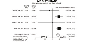
Figure 1. New meta-analysis graphic
In conclusion, establishing the 6th day as a routine for collecting free-DNA for niPGT-A will undoubtedly be the third disobedience of PGT regarding the principle of primum non nocere.
REFERENCES
Barash OO, Ivani KA, Willman SP, Rosenbluth EM, Wachs
DS, Hinckley MD, Pittenger Reid S, Weckstein LN. Association
between growth dynamics, morphological parameters,
the chromosomal status of the blastocysts, and clinical outcomes in IVF PGS cycles with single embryo transfer.
J Assist Reprod Genet 2017;34:1007-6. PMID: 28560610
DOI: 10.1007/s10815-017-0944-0
Medline Crossref
Bourdon M, Pocate-Cheriet K, Finet de Bantel A, Grzegorczyk-
Martin V, Amar Hoffet A, Arbo E, Poulain M, Santulli
P. Day 5 versus Day 6 blastocyst transfers: a systematic
review and meta-analysis of clinical outcomes.
Hum Reprod. 2019; 34:1948-64. PMID: 31644803. DOI:
10.1093/humrep/dez163.
Medline Crossref
Coates A, Kung A, Mounts E, Hesla J, Bankowski B, Barbieri
E, Ata B, Cohen J, Munné S. Optimal euploid embryo
transfer strategy, fresh versus frozen, after preimplantation
genetic screening with next generation sequencing: a
randomized controlled trial. Fertil Steril 2017;107:723-30.
PMID: 28139240. DOI: 10.1016/j.fertnstert.2016.12.022.
Medline Crossref
Fang R, Yang W, Zhao X, Xiong F, Guo C, Xiao J, Chen L,
Song X, Wang H, Chen J, Xiao X, Yao B, Cai LY. Chromosome
screening using culture medium of embryos fertilised
in vitro: a pilot clinical study. J Transl Med. 2019;17:73.
PMID: 30849973 DOI: 10.1186/s12967-019-1827-1.
Medline Crossref
Greco E, Minasi MG, Fiorentino F. Healthy Babies after
Intrauterine Transfer of Mosaic Aneuploid Blastocysts. N
Engl J Med. 2015;373:2089-90. PMID: 26581010 DOI:
10.1056/NEJMc1500421.
Medline Crossref
Jiao J, Shi B, Sagnelli M, Yang D, Yao Y, Li W, Sha L, Lu S,
Li D, Wang X. Minimally invasive preimplantation genetic
testing using blastocyst culture medium. Hum Reprod
2019;34:1369-79. PMID: 31251795 DOI: 10.1093/humrep/
dez075
Medline Crossref
Irani M, O’Neill C, Palermo GD, Xu K, Zhang C, Qin X, Zhan
Q, Clarke RN, Ye Z, Zaninovic N, Rosenwaks Z. Blastocyst
development rate influences implantation and live birth
rates of similarly graded euploid blastocysts. Fertil Steril.
2018;110:95-102.e1. PMID: 29908774. DOI: 10.1016/j.
fertnstert.2018.03.032.
Medline Crossref
Mastenbroek S, Twist M, van Echten-Arends J, Sikke ma-Raddatz B, Korevaar JC, Verhoeve HR, Vogel NE, Arts
EG, de Vris JW, Bossuyt PM, Buys CH, Heineman MJ, Repping
S, van der Veen F. In vitro fertilization with preimplantation
genetic screening. N Engl J Med 2007; 357:9-17.
PMID: 17611204. DOI: 10.1056/NEJMoa067744
Medline Crossref
Popovic M, Dhaenens L, Boel A, Menten B, Heindryckx B.
Chromosomal mosaicism in human blastocysts: the ultimate
diagnostic dilemma. Hum Reprod Update. 2020; 26:
313-34. PMID: 32141501 DOI: 10.1093/humupd/dmz050.
Medline Crossref
Rubio C, Rienzi L, Navarro-Sánchez L, Cimadomo D,
García-Pascual CM, Albricci L, Soscia D, Valbuena D, Capalbo
A, Ubaldi F, Simón C. Embryonic cell-free DNA
versus trophec- toderm biopsy for aneuploidy testing:
concordance rate and clinical implications. Fertil Steril.
2019;112:510-9. PMID: 31200971 DOI: 10.1016/j.fertnstert.
2019.04.038.
Medline Crossref
Rubio C, Navarro-Sánchez L, García-Pascual CM, Ocali O,
Cimadomo D, Venier W, Barroso G, Kopcow L, Bahçeci M,
Kulmann MIR, López L, De la Fuente E, Navarro R, Valbuena
D, Sakkas D, Rienzi L, Simón C. Multicenter prospective
study of concordance between embryonic cell-free DNA
and trophectoderm biopsies from 1301 human blastocysts.
Am J Obstet Gynecol. 2020:S0002-9378(20)30520-2.
PMID: 32470458. DOI: 10.1016/j.ajog.2020.04.035.
Medline Crossref
Taylor TH, Patrick JL, Gitlin SA, Wilson JM, Crain JL, Griffin
DK. Comparison of aneuploidy, pregnancy and live
birth rates between day 5 and day 6 blastocysts. Reprod
Biomed Online. 2014;29:305-10. PMID: 25043891. DOI:
10.1016/j.rbmo.2014.06.001.
Medline Crossref
Vagnini LD, Petersen CG, Renzi A, Dieamant D, Oliveira
JBA, Oliani AH, Canas MCT, Nakano R, Almodin CG, Marcondes
C, Ceschin A, Amaral A, Soares JB, Lopes J, Franco
AC, Franco Jr JG. Relationship between age and blastocyst
chromosomal ploidy analysed by noninvasive preimplantation
genetic testing for aneuploidies (niPGT-A). JBRA Assist
Reprod. 2020. PMID: 32723707. DOI: 10.5935/1518-
0557.20200061.
Medline Crossref
Xu J, Fang R, Chen L, Chen D, Xiao JP, Yang W, Wang H,
Song X, Ma T, Bo S, Shi C, Ren J, L, Cai LY, Yao B, Xie XS, Lu
S. Noninvasive chromosome screening of human embryos
by genome sequencing of embryo culture medium for in vitro
fertilization. Proc Natl Acad Sci USA. 2016; 113:11907-
12. PMID: 2768876 DOI: 10.1073/pnas.1613294113.
Medline Crossref