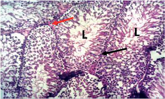
Figure 2. Photomicrograph of the testes from a tiger nut alone treated rat, showing typical seminiferous tubule, demonstrating the lumen (L) basement membrane (black arrow) and interstitial space (red arrow). H&E X100.
Figure 2.
Photomicrograph of the testes from a tiger nut alone treated rat, showing typical seminiferous tubule, demonstrating the lumen (L) basement membrane (black arrow) and interstitial space (red arrow). H&E X100.