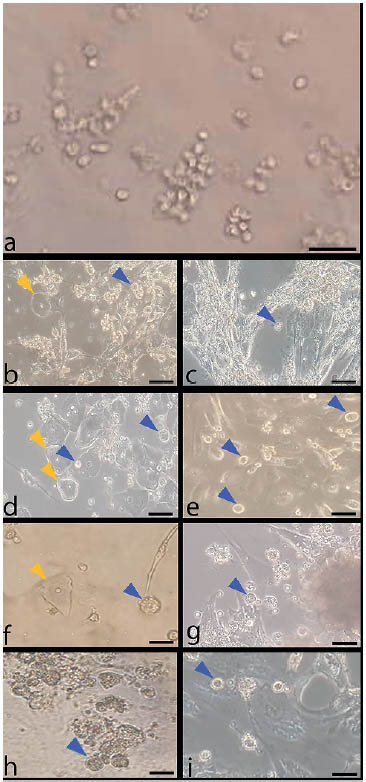
Figure 1. Morphological changes of follicular fluid cells in minimal culture condition. Control groups without melatonin treatment on day 0(a), week 1(b), week 2(c); groups treated with melatonin 10-7M on week 1(d) and week 2 (e); groups treated with melatonin 10-9M on week 1(f) and week 2(g); groups treated with melatonin 10-11M on week 1(h) and week 2(i); blue arrows: OLCs; yellow arrows: epithelial cells; 20 X.