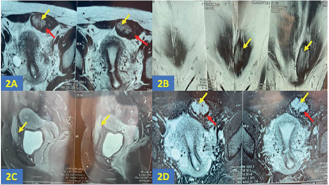
Figure 2. 2A (axial T2WI), 2B (coronal T2WI), 2C (T1WI-post contrast), 2D (STIR axial): the yellow arrows in these images reveal a relatively well defined nodular lesion in the supra-pubic region within the bulky left rectus abdominis muscle, measuring 5.8cm x 3.3cm x 2.5cm. The lesion appears heterogeneously hyperintense on T2WI and STIR, showing a peripheral hypointense rim due to hemosiderin deposition (red arrow- 2A, 2D) with post contrast enhancement (2C).