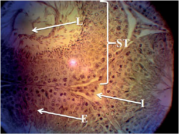
Figure 1. Histology of the testis of the control group showing normal testicular histology with intact seminiferous tubules (ST), interstitium containing leydig cell (I), seminiferous epithelium containing the spermatogenic cells and sertoli cells (E) and seminiferous lumen (L). H&E stain; (X400).