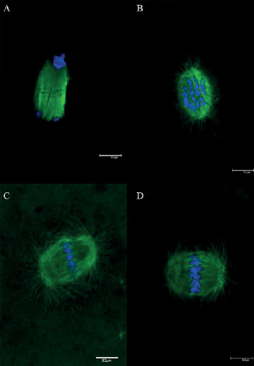
Figure 2. Nuclear maturation stages of murine oocytes matured in vivo and visualized by confocal microscopy. Note: (A) telophase I (TI) stage; (B) meiotic spindle viewed in a polar position (not analyzable); (C and D) meiotic spindle in a sagittal position (analyzable). Green: spindle marked with anti-β-tubulin antibody and secondary antibody conjugated with fluorescein isothiocyanate; blue: chromosomes marked with DAPI. Scale bar = 10 μm.