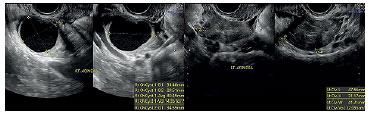
Figure 6. Pelvic ultrasound performed during the follicular phase three months after surgery. Despite of the ovary volumes achieved, only two and one antral follicles were displayed, on the left and right ovaries respectively. On the right ovary a cystic lesion could be seen. RT- right; LT- left.