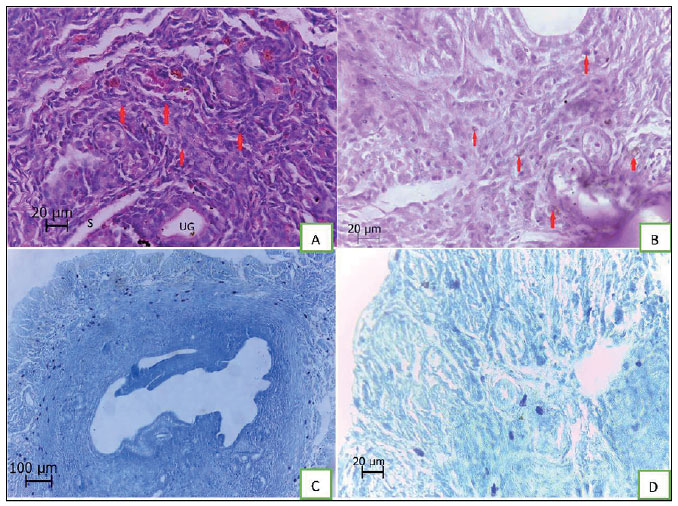
Figure 1. Morphology of uterus samples. Micrographs A: UG indicates uterine glands, S indicates sinusoid and the arrows indicate decidual cells (H&E, ×400). Micrographs B: decidualization of endometrium and the arrowed cells indicate large granular inflammatory cells (PAS, ×400). Micrographs C and D: The dark blue cells indicate mast cells (toluidine blue, ×100 [C] and ×400 [D]). Distribution of mast cells is seen that most of them are in myometrium.