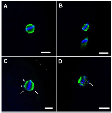
Figure 1. Representative confocal microscopy images (40x) of bovine oocytes matured in vitro during metaphase II, based on organization of the meiotic spindle and chromosomal alignment. Normal MII: A; Abnormal MII: B, C, D. Note: Scale bar: 10 µm; White arrows: misaligned chromosomes.