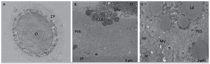
Figure 3. Microscopic and ultrastructural evaluation of an oocyte from patient 1. Coarse granules in the PVS can be clearly observed at 100X magnification with an inverted microscope (A). Lipid structures are observed either in association with microvilli (B) or dispersed within the PVS (C). O, oocyte; ZP, zona pellucida; PB, polar body; Mv, microvilli; PVS, perivitelline space; M, mitochondria; Ld, lipid droplet.