JBRA Assist. Reprod. 2023;27(4):668-676
ORIGINAL ARTICLE
doi: 10.5935/1518-0557.20230045
The effect of kisspeptin on the maturation of human ovarian follicles in culture following vitrification-thawing processes
1Men’s Health and Reproductive Health Research Center, Shahid Beheshti University of Medical Sciences, Tehran, Iran
2Department of Biology, Faculty of Science, Arak University, Arak, Iran
3Clinical Research Development Unit of Imam Khomeini Hospital, Urmia University of Medical Sciences, Urmia, Iran
4Department of Anatomy, School of Medicine, Fasa University of Medical Sciences, Fasa, Iran
5Department of obstetrics and gynecology, Atieh hospital, Tehran, Iran
6Department of Biology and Anatomical Sciences, School of Medicine, Shahid Beheshti University of Medical Sciences, Tehran, Iran
CONFLICT OF INTERESTS
The authors declare that they have no competing interests.
ABSTRACT
Objective: Ovarian cryopreservation is one of the effective methods to preserve fertility for cancer patients. Still, this approach has some problems, namely ROS, resulting in adverse effects on oocytes and ovarian follicles. Kisspeptin as an antioxidant to control ovarian function, directly or indirectly. In this study, the effect of kisspeptin on follicle maturation was evaluated in culture following ovarian cryopreservation.
Methods: Ovarian tissue samples of women between 20 and 35 years old (n=12) were laparoscopically collected. The samples were randomly divided into four groups: 1) control, 2) vitrification, 3) vitrified+1μM kisspeptin, and 4) vitrified+10μM kisspeptin. After vitrification and thawing processes, the tissues were cultured in DMEM medium for 7 days. H&E staining for histological evaluation, Real-Time PCR for GDF9 and BMP15 gene expression, and immunohistochemical staining for GDF9 and BMP15 protein expression were performed.
Results: In the vitrification group, ovarian tissue morphology was incoherent, and more primordial follicles than other follicle types were found. The expression of GDF9 and BMP15 genes and proteins were significantly decreased in this group compared with other groups (p<0.05). In the vitrification groups with kisspeptin (1 and 10 μM), the number of primary and secondary follicles was more than in the vitrification group. Besides, the expression of these genes and proteins was dramatically elevated in the vitrification groups with kisspeptin compared to the vitrification group alone (p<0.05).
Conclusions: It seems that kisspeptin is an effective substance to improve the quality of the human ovarian cryopreservation medium by improving follicle maturation.
Keywords: BMP15, follicle maturation, GDF 9, human ovarian cryopreservation, kisspeptin
INTRODUCTION
Yearly, a great number of reproductive-age adult women all over the world find out they have cancer. One of the main challenges for using common medical approaches for cancer treatment in women is maintaining women’s fertility, especially in underage girls (Rivas Leonel et al., 2019). Treatment methods for cancer, such as radiation therapy and chemotherapy, increase the survival rates of patients; however, these treatments can lead to POI (Primary ovarian insufficiency) and eventually pregnancy disruption (Blumenfeld, 2012; Kim et al., 2021). Even demands for fertility preservation for non-oncological reasons are increasing day by day (Donnez & Dolmans, 2017). Thus, finding effective techniques for fertility preservation in these cases is essential (Kim et al., 2021; Aliakbari et al., 2022).
Among this, ovarian cryopreservation has been introduced as a useful method for prepubertal girls with cancer, and patients who are unable to delay treatment or ovarian stimulation is harmful to them (Jadoul et al., 2010; Wallace et al., 2014). Recently, cryopreservation of ovarian tissue by vitrification has been considered an important tool for the preservation of female fertility, in particular for subjects undergoing chemotherapy (Yang et al., 2016). Vitrification is a more cost-effective and faster method than slow freezing and it also prevents the formation of ice crystals in cells, causing high rates of embryo development, cell survival, and fertilization (Shi et al., 2017; Huo et al., 2021). Also, thawing is known as a fast way to prevent the re-crystallization process in which water hits embryos or oocytes and can be transformed into a solid form around, creating small ice crystals (Chian et al., 2014). One of the main results of the vitrification and thawing processes is ROS (Reactive oxygen species) formation through various mechanisms, like osmotic stress, oxidative metabolism, and changes in cell defense mechanisms that can lead to apoptosis stimulation and subsequent DNA damage (Mazoochi et al., 2009; Tatone et al., 2010; Rocha et al., 2018).
In the women’s reproductive system, some agents suppress the apoptosis of granulosa cells and follicular atresia and stimulate granulosa cell mitosis, e.g., BMP15 (Bone morphogenetic protein 15) and GDF9 (Growth differentiation factor 9) (Gode et al., 2011). Several studies have been performed to decrease cryopreservation-stimulated cell injuries; however, there is no efficient method to date to treat these lesions (Rocha et al., 2018). In this way, kisspeptin (metastin), as a neuropeptide whose gene (KISS1) is located on chromosome 1q32.11, regulates the expression of antioxidant enzymes against oxidant agents and controls puberty and reproductive activities through its membrane receptors coupled with the G protein (Kotani et al., 2001). Kisspeptin triggers hypothalamic pituitary gonadal axis inducting gametogenesis by releasing FSH (Follicle stimulating hormone) and LH (Luteinizing hormone) through the pituitary gland (Aslan et al., 2017; MacManes et al., 2017). Also, reports expressed the role of kisspeptin in ovarian function control, such as follicular development, steroidogenesis, oocyte maturation, and ovulation (Hu et al., 2018). Despite these, according to our knowledge, the effectiveness of this neuropeptide on the maturity of human ovarian follicles in a vitrification medium has not been investigated. Hence, in this investigation, we studied the effect of kisspeptin in culture following vitrification-thawing methods on follicle maturation and the expression of related genes and proteins (BMP15 and GDF9) in vitro for the first time.
MATERIALS AND METHODS
Sample collection
This study has been approved by the Shahid Beheshti University of Medical Sciences (ethical code: IR.SBMU.RETECH.REC.1399.554). We used the ovarian tissues of 20 women between the ages of 20 and 35 who underwent a hysterectomy and had their fallopian tubes ligated. All the patients were informed about the study, and their consent was obtained. The health of human ovarian tissue was examined by an obstetrician, and only women with normal BMI (Body mass index) (<27kg/m2) (Diamanti-Kandarakis & Bergiele, 2001) and normal AMH levels (Anti-mullerian hormone) (1.66ng/ml) (Yang et al., 2017) were included in the project (Table 1). Exclusion criteria were: Ovaries damaged for any reason during surgery, etc., as well as cancerous ovaries, ovaries without normal follicles, ovaries of people undergoing chemotherapy or hormone therapy, ovaries of people with addiction, ovaries of people using corticosteroids, and polycystic ovaries (Hardy, 2018). A sample of each ovary was taken to check the quality of follicles histologically by H&E (Hematoxylin and eosin) staining. Finally, 12 ovaries were healthy and had normal follicles, and others were excluded. Table 1 depicts the characteristics of the subjects involved in the study are shown in Table 1.
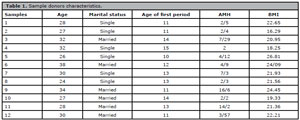
Table 1. Sample donors characteristics.
The tissues of those ovaries removed laparoscopically by the obstetrician were transferred to the laboratory in 10% HAMS (Ham’s tissue culture medium) solution and 20% human albumin serum with ice at -4°C for one h. The ovarian tissues were divided into 5 x 5 x 1 mm pieces with a sharp blade. All steps were performed on ice and under the laminar hood. Human ovarian cortex tissue parts were randomly allocated into four groups. All the mentioned chemicals were purchased from Sigma-Aldrich Chemie, Steinheim, Germany.
Experimental groups
From each one of 12 ovaries obtained we collected 12 samples. We assigned three ovarian samples from each one of 12 samples to each group and, eventually, each of the experimental groups was further divided into four groups:
I. Control group: Fresh ovarian samples (n=36)
II. Vitrification group: Ovarian samples underwent culture following vitrificationthawing methods (without adding Kisspeptin) (n=36)
III. Vitrification with + 1 µM kisspeptin group: 1 µM kisspeptin was added to vitrification medium and ovarian samples underwent vitrification-thawing methods (n=36)
IV. Vitrification + 10 µM kisspeptin group: 10 µM kisspeptin was added to vitrification medium and ovarian samples underwent vitrification-thawing methods (n=36)
Vitrification
The ovarian tissue samples were placed by a carrier for 25 min in an ES (Equilibration solution) medium containing 7.5% ethylene glycol and 7.5% DMSO (Dimethyl sulphoxide) in 10% HAMS as a handling medium and then followed by a second equilibration VS (Vitrification solution) for 15 min containing 20% ethylene glycol, 20% DMSO, and 0.5 mol/ L Sucrose with 10% HEPES (4-(2-hydroxyethyl)-1-piperazineethanesulfonic acid). Kisspeptin was added to the VS medium at the concentration of 1 µM for group III and 10 µM for group IV.
Thawing
For the thawing process, the ovarian samples were first immersed in a T₁ medium containing 1 mole sucrose and 10% HEPES with HAMS as the base medium for 1 min and then for 5 min in a T₂ medium containing 10% HEPES and 0.5 mol sucrose followed by putting them in a T₃ medium containing 10% HEPES and 0.25 mol sucrose for 10 min.
Tissue culture
After thawing, the ovarian samples were cultured for seven days. The medium was DMEM 12 (Dulbecco’s modified eagle medium) ready culture with 10% FBS (Fetal bovine serum), and 5% Streptomycin and Penicillin antibiotics were used in the incubator to maintain the tissue for different stages of the tests. The culture medium was changed every two days and it didn’t contain kisspeptin.
Histological assessments
All tissue samples after vitrification-thawing processes were fixed in 10% formalin. Tissue samples were embedded in paraffin blocks after tissue processing through graded alcohol and xylene solutions. Paraffin blocks were serially sectioned at a thickness of 5 µm. Afterwards, the sections were stained with H&E, mounted, and observed under light microscopy (Olympus, Tokyo, Japan). The identification and classification of follicles were done according to the study by Dehghani et al. (2018).
Quantitative real-time polymerase chain reaction
In all groups, 2 or 3 fragments were homogenized and total RNA was extracted (Parstous, Iran). Then, isolated RNAs were converted to cDNA by cDNA synthesis kit (Parstous, Iran). Appropriate primers for BMP15, GDF9, and GAPDH genes were designed using Gene Bank database (http://www.ncbi.nlm.nih.gov) and primer software (Blast, MEDUSA, Primer 3 and UCSC). The primer sequences, product length, and GenBank access numbers are shown on Table 2. The primers were ordered from and synthesized at Sinaclon Company, Iran. In this investigation, the master mix PCR from (Parstous, Iran) was used comprising 5µl 2χ Master Mix, 0.5 µl Forward primer (10 µM), 0.5 µl Reverse primer (10µM), and 3µl H₂O (Total Volume=9µl + 1µl cDNA). All PCR materials were prepared in a 0.2 ml microtube and mixed and spun. The final mixture was distributed in the volumes of 9 μl in PCR microtubes, and 1 μl cDNA or DNA was added to each microtube. Next, the microtubes were put in the Real-time PCR thermocycler (ABI Step One, USA) at 95 °C for 5 min (Initial denaturation), 95°C for 15 sec, 50-60°C for 20 sec, 72°C for 30 sec (Amplification) and 4°C for 5 min (cooling). To check the gene expression and the specificity of PCR products, agarose gel with a suitable percentage depending on the length of the PCR product was prepared, and all PCR products were electrophoresed.
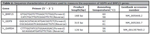
Table 2. Sequence characteristics of primers used to measure the expression of GDF9 and BMP15 genes.
Preparation of the electrophoresis gel
To prepare the 1% gel, 0.5 g agarose was mixed with 10 ml TBE buffer (Merck, Germany) in an Erlenmeyer flask, which was placed on heat until it became clear and reached a temperature of 70 degrees Celsius. Next, the special dye (safe stain Merck, Germany) was added to the mixture and poured into the electrophoresis tank. The gel was covered with 1% buffer. After the gel hardened, 3 μl of the product was combined with 1 μl buffer containing the dye (Merck, Germany) and inserted into the electrophoresis tank wells. A sizing solution (Sigma Aldrich, Germany) was poured into one of the wells, and the length of parts was determined accordingly. After entering the samples, the electrophoresis device was set to a voltage of 80 and a time of 30 min. After that, the gel was transferred to the screen of the trans luminaire device (Royan, Iran), and the band of samples was analyzed for the presence of the band, density, and dimensions (Figure 1).
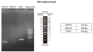
Figure 1. (A) shows ovarian tissue in the control group. In this group, more coherence was seen in the tissue than in other groups. (B) shows the H&E staining in the vitrification group and the primordial follicles can be seen. Also, tissue cohesion due to the vitrification and staining steps is less than in the control group. (C) indicates tissue incoherence, and primary follicles were seen in this group. (D) reveals the incoherence of primary and secondary tissues and Graafian follicles.
Immunohistochemistry
For IHC staining, paraffin-embedded ovarian tissue slides were deparaffinized with xylene and rehydrated with a series of ethanol solutions (50%, 70%, 80%, 90%, and 100%). 2 N hydrochloric acid was poured on the samples for 30 min, then borate buffer was added for 5 min. Tissue samples were washed with phosphate-buffered saline (PBS), and then 0.3% Triton was used for 30 min to permeate the cell membranes and washed with PBS. Afterward, 10% goat serum was added for 30 min as an additional background color. The primary antibody (GDF9-orb13431 and BMP15-orb247897) (1: 100) was diluted with PBS and added to the samples, then placed in a refrigerator at 2 to 8°C overnight after creating a humid environment to prevent tissue drying. After 24 h, the tissue container was removed from the refrigerator and then washed 4 times with PBS for 5 min each time. Secondary antibodies (orb688925) were added to the samples at a dilution of 1 to 150 and subsequently incubated at 37°C for 1 h and 30 min in the dark. The samples were transferred from the incubator to a dark room, and after 4 washings, DAPI (4′,6-diamidino-2-phenylindole) was added to them, immediately removed, and poured on the PBS samples. In the last step, the samples were studied in an Olympus fluorescent microscope (Olympus, Tokyo, Japan) (400 X) to confirm the markers.
Statistical analyses
The data were analyzed statistically by the GraphPad Prism software, version 8.4.3, using the one-way ANOVA for the differences between more than two groups and Tukey’s tests for differences between groups. The results were reported as mean ± SEM. The level of significance was considered at p<0.05.
RESULTS
Histological findings
In the qualitative investigation of human ovarian tissue, there was more tissue cohesion in the control group than in the other groups. Also, stroma cells, granulosa, and follicles revealed a regular structure in the normal group. In the vitrified group compared to the control group, tissue cohesion was less, and there were more primordial follicles than other follicle types. In this group, the order of granulosa cells and the structure of follicles at different stages of growth (except for primordial follicles) were disrupted. Also, the number of primary and secondary follicles in vitrified + 1 μM kisspeptin and vitrified + 10 μM kisspeptin groups was more than in the vitrification group. In the secondary follicles, the surrounding granulosa cells and the antrum cavity were visible. In the vitrified + 1 μM kisspeptin group and the vitrified + 10 μM kisspeptin group, the number of primordial follicles was reduced. In most follicles, the nuclei were damaged and not seen due to the staining processes (Figure 2).
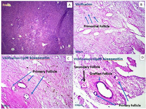
Figure 2. Gel electrophoresis of GDF9, BMP15, and GAPDH genes.
Gene expression
1- GDF9
In the vitrification group, the expression of the GDF9 gene was significantly reduced in comparison with other groups (p<0.05). Plus, the expression of this gene was elevated in the vitrified + 1 μM kisspeptin and vitrified + 10 μM kisspeptin groups than in the vitrified groups (p<0.05). In addition, gene expression of GDF9 in the vitrified + 10 μM kisspeptin group was higher than in the vitrified + 1 μM kisspeptin group (p<0.05). Also, there were no significant differences in the expression of the GDF9 gene between the vitrification + 1 μM kisspeptin and the control groups (p<0.05) (Figure 3).
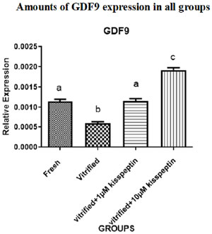
Figure 3. Expression of GDF9 gene in different groups after seven days of the vitrification and seven days of culture in DMEM medium. Data are shown as Means ± SD (One way ANOVA, Tukey´s test, p<0.05).
2- BMP15
BMP15 gene expression was significantly decreased in the vitrified group compared with other groups (p<0.05). The expression of this gene was also elevated in the vitrified + 1 μM kisspeptin and vitrified + 10 μM kisspeptin groups compared with the fresh and vitrified groups (p<0.05). In addition, gene expression of BMP15 in the vitrified + 10 μM kisspeptin group was higher than in the vitrified + 1 μM kisspeptin group (p<0.05) (Figure 4).
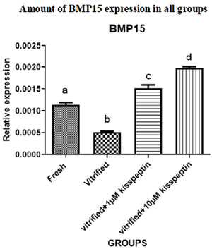
Figure 4. BMP15 gene expression after seven days of the glass freezing and seven days of culture in DMEM medium. Data are shown as Means ± SD (One way ANOVA, Tukey´s test, p<0.05).
Protein expression
3- GDF9
The protein expression of the GDF9 gene was dramatically diminished in the vitrified group compared with other groups (p<0.05). The protein expression of the mentioned gene in the vitrified + 1 μM kisspeptin and vitrified + 10 μM kisspeptin groups was significantly increased than in the vitrified group (p<0.05). Also, the protein expression of GDF9 in the vitrified + 10 μM kisspeptin group was higher than in the vitrified + 1 μM kisspeptin group (p<0.05) (Figures 5A and B).

Figure 5A. In A, B, C, and D, the green fluorescent color indicates the expression of GDF9 protein in the ovarian tissue in different groups after seven days of the vitrification and then seven days of culture in DMEM medium. In E, F, G, and H, the cell nuclei turned blue. In I, J, K, and L, the combination of the two above rows can be seen.

Figure 5B. evaluation of the average percentage of gdf9 protein expression in different groups after seven days of the vitrification and seven days of culture in dmem medium. data are shown as means ± SD (One way ANOVA, Tukey’s test, p<0.05).
4- BMP15
The protein expression of the BMP15 gene was significantly decreased in the vitrified group than in other groups (p<0.05). The protein expression of this gene was dramatically elevated in the vitrified + 1 μM kisspeptin and vitrified + 10 μM kisspeptin groups compared with the vitrified group (p<0.05). Plus, the protein expression of BMP15 in the vitrified + 10 μM kisspeptin group was higher than in the vitrified + 1 μM kisspeptin group (p<0.05) (Figures 6A and B).

Figure 6A. In A, B, C, and D, the green fluorescent color indicates the expression of BMP15 protein in the ovarian tissue in different groups. In E, F, G, and H, the nuclei of normal cells turned blue. Also, in I, J, K, and L, the combination of the first and second rows can be seen.

Figure 6B. Evaluation of the average percentage of BMP15 protein expression in different groups after seven days of the vitrification and then seven days of culture in DMEM medium. Data are shown as Means ± SD (One way ANOVA, Tukey´s test, p<0.05).
DISCUSSION
Ovarian tissue cryopreservation through vitrification and thawing techniques is a potential tool to preserve fertility in women who suffer from cancer. However, this method is accompanied by ROS production and subsequently DNA damage, which can have detrimental effects on the oocytes and ovarian follicles (Tsai-Turton & Luderer, 2006; Silva et al., 2018; Tamura et al., 2020). The optimization of this approach has not been carried out yet (Nyachieo et al., 2013; Dolmans & Donnez, 2021).
On the other hand, using antioxidants has been recommended as a practical way to minimize the production of ROS during cryopreservation (Silva et al., 2018). In this regard, we evaluated the effectiveness of kisspeptin, as an antioxidant agent (Aslan et al., 2017), on follicle maturation and the expression of relevant genes and proteins (BMP15 and GDF9) based on ovarian tissue culture following vitrification-thawing processes.
In this study, the histological results revealed a reduction in the number of follicles in all growth stages with the exception of primordial follicles in the vitrification group than in the fresh group. These findings may reflect the negative effects of the vitrification on follicle maturation in light of ROS formation (Chang et al., 2019; Shoorei et al., 2019). Similarly, Migishima et al. (2003) indicated a decrease in the number of follicles in frozen-thawed ovaries than in fresh ovaries. However, some documents reported normal morphology of ovarian follicles after the vitrification (Salehnia et al., 2002; Luvoni et al., 2012). We also observed that the number of primary and secondary follicles in vitrification groups with kisspeptin (1 and 10 μM) was more than in the vitrification group.
It has been declared that kisspeptin potentiates the induction of ovarian follicle maturation and estradiol production by promoting FSH synthesis (Hagen et al., 2014; Rather et al., 2016). The line with this notion, some published papers emphasized the role of this antioxidant in improving follicle development and maturity (Taniguchi et al., 2017; Magamage et al., 2021). In this regard, a study revealed that the administration of kisspeptin increases the number of preovulatory follicles and raises the plasma AMH secreted by secondary follicles (Cao et al., 2019). In this study, the gene and protein expression of GDF9 and BMP15, as follicle maturity agents, in the vitrification group was significantly reduced compared with the control group. Consistent with this finding, Choi et al. (2007) showed diminished expression of some markers related to follicle development, like GDF9, after cryopreservation of mouse ovaries. Also, Ebrahimi et al. (2010) reported that the expression of GDF9 and BMP15 was decreased owing to freezing sheep oocytes. Unlike these studies, some researches manifested that the gene expression of GDF9 did not have a significant difference compared to the control group after freezing (Ramezani et al., 2017). These differences may be related to differences in cryopreservation methods (Stachowiak et al., 2009). Another report was that the gene and protein expression of GDF9 and BMP15 in the vitrification groups with kisspeptin was significantly elevated compared with the vitrification group. In corresponding with this result, Saadeldin et al. (2012) demonstrated increased expression of GDF9 and BMP15 genes in the time of in vitro maturation of oocytes after kisspeptin administration. Also, it is stated that oocyte maturation stimulated by kisspeptin is in light of GDF9 and BMP15 upregulation (Hu et al., 2018). BMP15 and GDF9 are crucial growth factors for regulating luteinization, folliculogenesis, ovulation, developmental competency, and oocyte maturation (Hu et al., 2018). In addition, their proteins are expressed in the primary stage of follicles and play an important role in the transfer of follicles from this stage to the secondary (Kona et al., 2016; Celik et al., 2020).
It looks like kisspeptin can serve as an obstacle against the destructive effects of the vitrification on follicle growth and maturation by elevating antioxidant defense processes and upregulating follicle maturity genes, like GDF9 and BMP15.
Limitation
In this study, due to the observance of ethical protocols, the number of samples was small, and less than 10% of the patient’s tissue was removed, so we were not able to perform further tests.
CONCLUSION
Our findings indicated that kisspeptin is a suitable substance to potentiate the quality of the human ovarian cryopreservation medium. Indeed, kisspeptin can increase the maturity of follicles and is effective to increase the possibility of a successful pregnancy using assisted reproductive techniques in clinics.
Abbreviations
POI, Primary ovarian insufficiency; OTC, Ovarian tissue cryopreservation; ROS, Reactive oxygen species; HAMs, Ham’s tissue culture medium; DMSO, Dimethyl sulphoxide; HEPES, 4-(2-hydroxyethyl)-1-piperazineethanesulfonic acid; ES, Equilibration solution; VS, Vitrification solution; DMEM, Dulbecco’s modified eagle medium; FBS, Fetal bovine serum; IHC, Immunohistochemistry; DAPI, 4′,6-diamidino-2-phenylindole; GDF9, Growth differentiation factor 9; BMP15, Bone morphogenetic protein 15; FSH, Follicle stimulating hormone; LH, Luteinizing hormone; BMI, Body mass index; AMH, Anti-mullerian hormone; H&E, Hematoxylin and eosin.
REFERENCES
Aliakbari F, Taghizabet N, Azizi F, Rezaei-Tazangi F, Samadee Gelehkolaee K, Kharazinejad E. A review of methods for preserving male fertility. Zygote. 2022;30:289-97. PMID: 34676815 DOI: 10.1017/S0967199421000071 Medline
Aslan M, Erkanli Senturk G, Akkaya H, Sahin S, Yılmaz B. The effect of oxytocin and Kisspeptin-10 in ovary and uterus of ischemia-reperfusion injured rats. Taiwan J Obstet Gynecol. 2017;56:456-62. PMID: 28805600 DOI: 10.1016/j.tjog.2016.12.018 Medline
Blumenfeld Z. Chemotherapy and fertility. Best Pract Res Clin Obstet Gynaecol. 2012;26:379-90. PMID: 22281514 DOI: 10.1016/j.bpobgyn.2011.11.008 Medline
Cao Y, Li Z, Jiang W, Ling Y, Kuang H. Reproductive functions of Kisspeptin/KISS1R Systems in the Periphery. Reprod Biol Endocrinol. 2019;17:65. PMID: 31399145 DOI: 10.1186/s12958-019-0511-x Medline
Celik S, Ozkavukcu S, Celik-Ozenci C. Altered expression of activator proteins that control follicle reserve after ovarian tissue cryopreservation/transplantation and primordial follicle loss prevention by rapamycin. J Assist Reprod Genet. 2020;37:2119-36. PMID: 32651677 DOI: 10.1007/s10815-020-01875-7 Medline
Chang H, Chen H, Zhang L, Wang Y, Xie X, Zhang Y, Quan F. Effect of oocyte vitrification on DNA damage in metaphase II oocytes and the resulting preimplantation embryos. Mol Reprod Dev. 2019;86:1603-14. PMID: 31408251 DOI: 10.1002/mrd.23247 Medline
Chian RC, Wang Y, Li YR. Oocyte vitrification: advances, progress and future goals. J Assist Reprod Genet. 2014;31:411-20. PMID: 24477781 DOI: 10.1007/s10815-014-0180-9 Medline
Choi J, Lee JY, Lee E, Yoon BK, Bae D, Choi D. Cryopreservation of the mouse ovary inhibits the onset of primordial follicle development. Cryobiology. 2007;54:55-62. PMID: 17196581 DOI: 10.1016/j.cryobiol.2006.11.003 Medline
Dehghani F, Aboutalebi H, Esmaeilpour T, Panjehshahin MR, Bordbar H. Effect of platelet-rich plasma (PRP) on ovarian structures in cyclophosphamide-induced ovarian failure in female rats: a stereological study. Toxicol Mech Methods. 2018;28:653-9. PMID: 29968488 DOI: 10.1080/15376516.2018.1491662 Medline
Diamanti-Kandarakis E, Bergiele A. The influence of obesity on hyperandrogenism and infertility in the female. Obes Rev. 2001;2:231-8. PMID: 12119994 DOI: 10.1046/j.1467-789x.2001.00041.x Medline
Dolmans MM, Donnez J. Fertility preservation in women for medical and social reasons: Oocytes vs ovarian tissue. Best Pract Res Clin Obstet Gynaecol. 2021;70:63-80. PMID: 32800711 DOI: 10.1016/j.bpobgyn.2020.06.011 Medline
Donnez J, Dolmans MM. Fertility Preservation in Women. N Engl J Med. 2017; 377:1657-65. PMID: 29069558 DOI: 10.1056/NEJMra1614676 Medline
Ebrahimi B, Valojerdi MR, Eftekhari-Yazdi P, Baharvand H, Farrokhi A. IVM and gene expression of sheep cumulus-oocyte complexes following different methods of vitrification. Reprod Biomed Online. 2010;20:26-34. PMID: 20158984 DOI: 10.1016/j.rbmo.2009.10.020 Medline
Gode F, Gulekli B, Dogan E, Korhan P, Dogan S, Bige O, Cimrin D, Atabey N. Influence of follicular fluid GDF9 and BMP15 on embryo quality. Fertil Steril. 2011;95:2274-8. PMID: 21496799 DOI: 10.1016/j.fertnstert.2011.03.045 Medline
Hagen CP, Sørensen K, Aksglaede L, Mouritsen A, Mieritz MG, Tinggaard J, Wohlfart-Veje C, Petersen JH, Main KM, Rajpert-De Meyts E, Almstrup K, Juul A. Pubertal onset in girls is strongly influenced by genetic variation affecting FSH action. Sci Rep. 2014;4:6412. PMID: 25231187 DOI: 10.1038/srep06412 Medline
Hu KL, Zhao H, Chang HM, Yu Y, Qiao J. Kisspeptin/Kisspeptin Receptor System in the Ovary. Front Endocrinol. 2018;8:365. PMID: 29354093 DOI: 10.3389/ fendo.2017.00365 Medline
Huo Y, Yuan P, Qin Q, Yan Z, Yan L, Liu P, Li R, Yan J, Qiao J. Effects of vitrification and cryostorage duration on single-cell RNA-Seq profiling of vitrified-thawed human metaphase II oocytes. Front Med. 2021;15:144-54. PMID: 32876878 DOI: 10.1007/s11684-020-0792-7 Medline
Jadoul P, Dolmans MM, Donnez J. Fertility preservation in girls during childhood: is it feasible, efficient and safe and to whom should it be proposed? Hum Reprod Update. 2010;16:617-30. PMID: 20462941 DOI: 10.1093/humupd/dmq010 Medline
Kim JM, Kim S, Lee S. Role of Stem Cells in the Ovarian Tissue Cryopreservation and Transplantation for Fertility Preservation. Int J Mol Sci. 2021;22:12482. DOI: 10.3390/ijms222212482 PMID: 34830363 Medline
Kona SS, Praveen Chakravarthi V, Siva Kumar AV, Srividya D, Padmaja K, Rao VH. Quantitative expression patterns of GDF9 and BMP15 genes in sheep ovarian follicles grown in vivo or cultured in vitro. Theriogenology. 2016;85:315-22. PMID: 26474685 DOI: 10.1016/j.theriogenology.2015.09.022 Medline
Kotani M, Detheux M, Vandenbogaerde A, Communi D, Vanderwinden JM, Le Poul E, Brézillon S, Tyldesley R, Suarez-Huerta N, Vandeput F, Blanpain C, Schiffmann SN, Vassart G, Parmentier M. The metastasis suppressor gene KiSS-1 encodes kisspeptins, the natural ligands of the orphan G protein-coupled receptor GPR54. J Biol Chem. 2001;276:34631-6. PMID: 11457843 DOI: 10.1074/jbc.M104847200 Medline
Luvoni GC, Tessaro I, Apparício M, Ruggeri E, Luciano AM, Modina SC. Effect of vitrification of feline ovarian cortex on follicular and oocyte quality and competence. Reprod Domest Anim. 2012;47:385-91. PMID: 21950518 DOI: 10.1111/j.1439-0531.2011.01885.x Medline
MacManes MD, Austin SH, Lang AS, Booth A, Farrar V, Calisi RM. Widespread patterns of sexually dimorphic gene expression in an avian hypothalamic-pituitary-gonadal (HPG) axis. Sci Rep. 2017;7:45125. PMID: 28417958 DOI: 10.1038/srep45125 Medline
Mazoochi T, Salehnia M, Pourbeiranvand S, Forouzandeh M, Mowla SJ, Hajizadeh E. Analysis of apoptosis and expression of genes related to apoptosis in cultures of follicles derived from vitrified and non-vitrified ovaries. Mol Hum Reprod. 2009;15:155-64. PMID: 19155222 DOI: 10.1093/molehr/gap002 Medline
Migishima F, Suzuki-Migishima R, Song SY, Kuramochi T, Azuma S, Nishijima M, Yokoyama M. Successful cryopreservation of mouse ovaries by vitrification. Biol Reprod. 2003;68:881-7. PMID: 12604638 DOI: 10.1095/biolreprod.102.007948 Medline
Nyachieo A, Spiessens C, Chai DC, Kiulia NM, Willemen D, Mwenda JM, Bourgain C, D’Hooghe TM. Ovarian tissue cryopreservation by vitrification in olive baboons (papio anubis): a pilot study. Gynecol Obstet Invest. 2013;75:157-62. PMID: 23391858 DOI: 10.1159/000346084 Medline
Ramezani M, Salehnia M, Jafarabadi M. Short Term Culture of Vitrified Human Ovarian Cortical Tissue to Assess the Cryopreservation Outcome: Molecular and Morphological Analysis. J Reprod Infertil. 2017;18:162-71. PMID: 28377895 Medline
Rather MA, Bhat IA, Sharma N, Sharma R, Gireesh-Babu P, Chaudhari A, Sundaray JK. Molecular characterization, tissue distribution of follicle-Stimulating Hormone (FSH) beta subunit and effect of kisspeptin-10 on reproductive hormonal profile of Catla catla (Hamilton, 1822). Aquacult Res. 2016;47:2089-100. PMID: 27088601 DOI: 10.1016/j.domaniend.2016.01.005 Medline
Rivas Leonel EC, Lucci CM, Amorim CA. Cryopreservation of Human Ovarian Tissue: A Review. Transfus Med Hemother. 2019;46:173-81. PMID: 31244585 DOI: 10.1159/000499054 Medline
Rocha CD, Soares MM, de Cássia Antonino D, Júnior JM, Freitas Mohallem RF, Ribeiro Rodrigues AP, Figueiredo JR, Beletti ME, Jacomini JO, Alves BG, Alves KA. Positive effect of resveratrol against preantral follicles degeneration after ovarian tissue vitrification. Theriogenology. 2018;114:244-51. PMID: 29660627 DOI: 10.1016/j.theriogenology.2018.04.004 Medline
Saadeldin IM, Koo OJ, Kang JT, Kwon DK, Park SJ, Kim SJ, Moon JH, Oh HJ, Jang G, Lee BC. Paradoxical effects of kisspeptin: it enhances oocyte in vitro maturation but has an adverse impact on hatched blastocysts during in vitro culture. Reprod Fertil Dev. 2012;24:656-68. PMID: 22697116 DOI: 10.1071/RD11118 Medline
Salehnia M, Abbasian Moghadam E, Rezazadeh Velojerdi M. Ultrastructure of follicles after vitrification of mouse ovarian tissue. Fertil Steril. 2002;78:644-5. PMID: 12215351 DOI: 10.1016/s0015-0282(02)03287-9 Medline
Shi Q, Xie Y, Wang Y, Li S. Vitrification versus slow freezing for human ovarian tissue cryopreservation: a systematic review and meta-anlaysis. Sci Rep. 2017;7:8538. PMID: 28819292 DOI: 10.1038/s41598-017-09005-7 Medline
Shoorei H, Banimohammad M, Kebria MM, Afshar M, Taheri MM, Shokoohi M, Farashah MS, Eftekharzadeh M, Akhiani O, Gaspar R, Pazoki-Toroudi H. Hesperidin improves the follicular development in 3D culture of isolated preantral ovarian follicles of mice. Exp Biol Med (Maywood). 2019;244:352-61. PMID: 30781997 DOI: 10.1177/1535370219831615 Medline
Silva LM, Mbemya GT, Guerreiro DD, Brito DCC, Donfack NJ, Morais MLGS, Rodrigues GQ, Bruno JB, Rocha RMP, Alves BG, Apgar GA, Cibin FWS, Figueiredo JR, Rodrigues APR. Effect of Catalase or Alpha Lipoic Acid Supplementation in the Vitrification Solution of Ovine Ovarian Tissue. Biopreserv Biobank. 2018;16:258-69. PMID: 29957024 DOI: 10.1089/bio.2017.0122 Medline
Stachowiak EM, Papis K, Kruszewski M, Iwaneńko T, Bartłomiejczyk T, Modliński JA. Comparison of the level(s) of DNA damage using Comet assay in bovine oocytes subjected to selected vitrification methods. Reprod Domest Anim. 2009;44:653-8. PMID: 19032440 DOI: 10.1111/j.1439-0531.2007.01042.x Medline
Tamura H, Jozaki M, Tanabe M, Shirafuta Y, Mihara Y, Shinagawa M, Tamura I, Maekawa R, Sato S, Taketani T, Takasaki A, Reiter RJ, Sugino N. Importance of Melatonin in Assisted Reproductive Technology and Ovarian Aging. Int J Mol Sci. 2020;21:1135. PMID: 32046301 DOI: 10.3390/ijms21031135 Medline
Taniguchi Y, Kuwahara A, Tachibana A, Yano Y, Yano K, Yamamoto Y, Yamasaki M, Iwasa T, Hinokio K, Matsuzaki T, Irahara M. Intra-follicular kisspeptin levels are related to oocyte maturation and gonadal hormones in patients who are undergoing assisted reproductive technology. Reprod Med Biol. 2017;16:380-5. PMID: 29259492 DOI: 10.1002/rmb2.12056 Medline
Tatone C, Di Emidio G, Vento M, Ciriminna R, Artini PG. Cryopreservation and oxidative stress in reproductive cells. Gynecol Endocrinol. 2010;26:563-7. PMID: 20230330 DOI: 10.3109/09513591003686395 Medline
Tsai-Turton M, Luderer U. Opposing effects of glutathione depletion and follicle-stimulating hormone on reactive oxygen species and apoptosis in cultured preovulatory rat follicles. Endocrinology. 2006;147:1224-36. PMID: 16339198 DOI: 10.1210/en.2005-1281 Medline
Wallace WH, Smith AG, Kelsey TW, Edgar AE, Anderson RA. Fertility preservation for girls and young women with cancer: population-based validation of criteria for ovarian tissue cryopreservation. Lancet Oncol. 2014;15:1129-36. PMID: 25130994 DOI: 10.1016/S1470-2045(14)70334-1 Medline
Yang MY, Cushman RA, Fortune JE. Anti-Müllerian hormone inhibits activation and growth of bovine ovarian follicles in vitro and is localized to growing follicles. Mol Hum Reprod. 2017;23:282-91. PMID: 28333275 DOI: 10.1093/molehr/gax010 Medline
Yang Y, Cheung HH, Law WN, Zhang C, Chan WY, Pei X, Wang Y. New Insights into the Role of Autophagy in Ovarian Cryopreservation by Vitrification. Biol Reprod. 2016;94:137. PMID: 26911431 DOI: 10.1095/biolreprod.115.136374 Medline