JBRA Assist. Reprod. 2024;28(2):299-305
REVIEW
doi: 10.5935/1518-0557.20230059
Antimüllerian hormone levels and IVF outcomes in polycystic ovary syndrome women: a scoping review
1Clínica Originare, Medicina Reprodutiva, São Paulo, Brazil
CONFLICT OF INTERESTS
The authors have no conflicts of interest to report.
ABSTRACT
Antimüllerian hormone (AMH) is a homodimeric glycoprotein secreted by granulosa cells from primary to large antral follicles, and it plays an important role in the regulation of early follicle growth. It is considered a reliable marker of ovarian reserve and a predictor of ovarian response to controlled stimulation. Polycystic ovary syndrome (PCOS) is an endocrine condition that affects women of reproductive age worldwide, and it is associated with high levels of AMH. PCOS patients may have worse maturation and fertilization rates compared to normo-ovulatory women. Some studies have demonstrated a positive correlation between AMH levels and qualitative aspects of assisted reproduction treatment; but it is not clear whether high levels of both serum and follicular fluid AMH in PCOS patients correlate with in vitro fertilization outcomes. We ran this scoping review of the literature to address this specific question. We comprehensively searched the databases PubMed and Cochrane Library until January 2023. We found that higher AMH levels are associated with higher oocyte yield, but PCOS patients tend to have fewer mature oocytes and impaired embryo quality and implantation rates. Pregnancy rates, however, are not affected by AMH levels or laboratorial outcomes. We also found that higher AMH levels are associated with worse PCOS features.
Keywords: polycystic ovary syndrome, antimüllerian hormone, in vitro fertilization, oocyte quality
INTRODUCTION
Antimüllerian hormone (AMH) is a homodimeric glycoprotein from the transforming growth factor-β superfamily secreted by granulosa cells from primary to large antral follicles (Desforges-Bullet et al., 2010; Mashiach et al., 2010; Pabuccu et al., 2009). It is suggested that AMH plays an important role in the regulation of early follicle growth, with an inhibiting effect on growing follicles (Mashiach et al., 2010), and suppressing the cyclical recruitment of primordial follicles (Tal et al., 2014).
AMH serum levels peak around 24 years of age and decline progressively until menopause (Melado Vidales et al., 2017a). It is considered a reliable marker of ovarian reserve and a predictor of ovarian response to controlled stimulation (Melado Vidales et al., 2017a; Tal et al., 2014). Some authors have suggested a positive correlation between AMH and oocyte quality, fertilization rates and embryo morphology (Borges et al., 2017; La Marca et al., 2011; Morales et al., 2022).
Polycystic ovary syndrome (PCOS) is an endocrine condition that affects 5-8% of women of reproductive age worldwide (Desforges-Bullet et al., 2010; Kaya et al., 2010). It is the most common cause of anovulatory infertility, and it is associated with high levels of AMH, which is a consequence, not only related to the higher number of preantral and small antral follicles, but also to an increased production by individual follicles (Kaya et al., 2010; Tal et al., 2014).
In PCOS patients, the selection of one follicle from the increased follicular pool and its posterior dominance is impaired. The mechanisms underlying this process are still uncertain, but inhibition of the local effect of follicle-stimulating hormone (FSH) seems to play an important role. AMH inhibits the initiation of primordial follicle growth by reducing the sensitivity of follicles to FSH, thus preventing the selection of multiple follicles (Arabzadeh et al., 2010). Thus, some studies have suggested that AMH plays an important role in the pathophysiology of PCOS. Serum AMH levels also appears to be related to the severity of PCOS and ovarian dysfunction, correlating with de degree of hyperandrogenism, ovulation disorder and polycystic ovary morphology (Tal et al., 2014).
Although some studies have demonstrated a positive correlation between AMH levels and qualitative aspects of assisted reproduction treatment, it is known that PCOS patients may have worse maturation and fertilization rates compared to normo-ovulatory women (Fallat et al., 1997; La Marca et al., 2011; Mashiach et al., 2010). The follicular microenvironment is also likely to be related to the success or failure of developmental competence of oocytes and embryos (Mashiach et al., 2010). Fallat et al. (1997) demonstrated that women with PCOS had higher follicular fluid AMH levels, a higher percentage of immature oocytes, and lower fertilization rates when compared to women with endometriosis and pelvic adhesions.
It is still controversial whether high levels of both serum and follicular fluid AMH in PCOS patients can correlate well with in vitro fertilization outcomes such as oocyte quality, fertilization rates and pregnancy rates or if AMH levels are restricted to the prediction of ovarian reserve in these patients. Thus, the aim of the present study is to perform a literature review regarding the association between the high levels of AMH and IVF outcomes in this specific population.
MATERIALS AND METHODS
We ran a literature review according to the PRISMA guideline (Preferred Reporting Items for Systematic Reviews and Meta-Analysis) to evaluate the study question. To identify the relevant studies, we searched the MEDLINE and Cochrane Library databases, with the last search made in January 2023 and no restriction regarding language or year of publication. Bibliographies of relevant studies identified by the search strategy and relevant reviews/meta-analyses were also searched for identification of additional studies. We retrieved the relevant articles using the search strategies described below:
MEDLINE: (((antimullerian hormone) AND (oocyte quality)) AND (embryo quality)) AND (polycystic ovary syndrome)
Cochrane Library: antimullerian hormone AND oocyte quality AND embryo quality AND polycystic ovary syndrome
The selection of manuscripts, as well as the evaluation of titles and abstracts, were conducted by two blinded researchers (L.C.D. and C.R.I) working independently. After this initial screening, the remaining articles were further evaluated to be included or not in the review.
RESULTS
The search process to identify and select the studies is presented in Figure 1. We found a total of 31 studies, of which 12 studies were included in the final review. Eighteen studies were excluded after an initial screening based on the titles. Four other studies were excluded after abstract reading for not individualizing patients with PCOS. Two other studies were excluded after full text reading because they were written in Chinese and no English text version could be retrieved. Five more studies were included after we searched the References of the studies already included. Table 1 summarizes the studies included in the review.
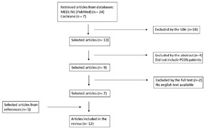
Figure 1. Flowchart of the studies included.
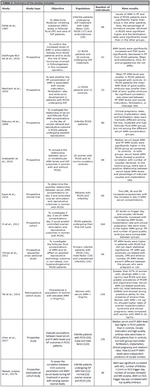
Table 1. Summary of the studies included.
Most of the studies were prospective, while only one was a retrospective cohort study. Eight of the twelve studies reported the follicular fluid levels of AMH (FF AMH). Seven of these eight studies compared PCOS patients with control normo-ovulatory patients. Five of them reported higher FF AMH in PCOS patients, while one study found lower FF AMH in PCOS patients, and another study did not find differences in FF AMH (Arabzadeh et al., 2010; Chen et al., 2017; Fallat et al., 1997; Mashiach et al., 2010; Melado Vidales et al., 2017b; Pabuccu et al., 2009; Yilmaz et al., 2012).
Seven studies reported the number of oocytes retrieved (Arabzadeh et al., 2010; Chen et al., 2017; Fallat et al., 1997; Mashiach et al., 2010; Melado Vidales et al., 2017a; Yilmaz et al., 2012, Xi et al., 2012). Fallat et al. (1997) found a higher number of immature oocytes in PCOS patients compared to patients with tubal factor infertility and endometriosis. Overall, there is a positive correlation between AMH levels and the number of oocytes retrieved in all patients, but not necessarily for the number of mature oocytes (Fallat et al., 1997). Mashiach et al. (2010) found no correlation between FF AMH and oocyte maturation in PCOS patients compared to controls. Arabzadeh et al. (2010) showed that higher AMH levels were associated with higher oocyte yield, but a positive relationship between serum AMH levels and mature oocytes was observed only in the non-PCOS group. Chen et al. also reported a lower oocyte maturation rate in PCOS patients (Chen et al., 2017).
Eight studies analyzed fertilization rates (FR), five of them comparing PCOS patients to normo-ovulatory patients, while the remaining three studies addressed PCOS patients only (Chen et al., 2017; Fallat et al., 1997; Kaya et al., 2010; Mashiach et al., 2010; Melado Vidales et al., 2017b; Pabuccu et al., 2009; Yilmaz et al., 2012; Xi et al., 2012). Fallat et al. (1997) were the only authors that found significantly lower fertilization rates in PCOS patients compared to controls. Pabuccu et al. (2009) showed that in PCOS patients the FR increased with higher FF AMH levels but did not correlate with serum AMH levels. Kaya et al. (2010) on the other hand, demonstrated a progressive increase in FR with higher serum AMH concentrations.
Five studies addressed the embryo quality and only one of them found PCOS patients had lower rates of high-quality embryos (Chen et al., 2017; Mashiach et al., 2010; Melado Vidales et al., 2017a; 2017b; Xi et al., 2012). Melado Vidales et al. (2017a) demonstrated serum AMH levels on hCG trigger day were correlated with the number of top-quality embryos obtained in all the patients studied. The same authors, however, found no significant between-group differences for AMH levels in FF nor for FR, number of TQE or implantation rates when comparing PCOS patients to patients with low/normal ovarian reserve (Melado Vidales et al., 2017b).
Six studies reported implantation rates (IR) and the results were discrepant (Arabzadeh et al., 2010; Chen et al., 2017; Kaya et al., 2010; Melado Vidales et al., 2017b; Pabuccu et al., 2009; Xi et al., 2012). The two studies that compared PCOS patients with normo-ovulatory controls did not find significant differences in IR between groups (Chen et al., 2017; Melado Vidales et al., 2017b). Of the studies that addressed only PCOS patients, Pabuccu et al. (2009) found IR were markedly increased with higher follicular-fluid AMH but not among the different serum AMH concentration groups. On the other hand, Kaya et al. (2010) showed IR increased progressively with the increase in day-3 AMH serum concentrations while Xi et al. (2012) found lower IR with higher serum AMH levels.
Clinical pregnancy rates (CPR) were reported in five of the twelve studies (Chen et al., 2017; Kaya et al., 2010; Pabuccu et al., 2009; Tal et al., 2014; Xi et al., 2012). Only one of them compared PCOS patients with normo-ovulatory controls and found similar rates, although the FF AMH levels were markedly higher in PCOS patients (Chen et al., 2017). Xi et al. (2012) and Kaya et al. (2010) studied only PCOS patients and showed conflicting data. While the first group of authors found CPR were lower in patients with higher serum AMH levels, the former demonstrated the opposite. Pabuccu et al. (2009) on the other hand, showed a positive correlation between CPR and FF AMH, but not serum AMH concentrations. A retrospective cohort study evaluated patients with serum AMH levels higher than 10 ng/mL and demonstrated a 97% prevalence of PCOS in this population. Moreover, these patients had a significantly higher CPR compared with patients who have AMH levels between 5 and 10 ng/mL (Tal et al., 2014).
DISCUSSION
The present study consisted of a scoping review that evaluated the association between AMH levels and IVF outcomes in women with infertility and PCOS. Our study showed that AMH levels in both serum and FF are higher in PCOS patients compared to women with other infertility factors. Regarding the laboratorial and pregnancy outcomes in IVF treatment, the studies analyzed were unanimous in concluding that PCOS women have a higher oocyte yield; however, whether this translates into better maturation, fertilization, implantation, clinical pregnancies, and live birth rates is still controversial.
AMH is a well-known marker of ovarian reserve and has been proven to be positively associated with ovarian response in controlled stimulation and, therefore, oocyte yield (Brodin et al., 2013). In PCOS the serum AMH levels are up to 5-fold higher than in normo-ovulatory women (Xi et al., 2012), mainly due to an increased in AMH production by each individual follicle rather than a higher number of follicles (Fanchin et al., 2005). In the past years, some studies have analyzed the AMH levels in follicular fluid during oocyte pickup (OPU) after controlled ovarian stimulation. As expected, most of them showed higher levels in PCOS patients (Arabzadeh et al., 2010; Chen et al., 2017; Fallat et al., 1997; Pabuccu et al., 2009; Yilmaz et al., 2012). Mashiach et al. (2010) found lower mean levels of AMH in FF of PCOS patients compared to normo-ovulatory controls; and Melado Vidales et al. (2017b) found no between-group differences when comparing PCOS women to patients with low and normal ovarian reserve. It is worth noting, though, that both studies are limited by small sample sizes. The AMH production by granulosa cells decreases during oocyte maturation, and the development of small antral follicles into mature follicles during controlled ovarian stimulation is associated with a dramatic decrease in serum AMH levels. Fanchin et al. (2005) demonstrated that FF AMH levels were 3 times higher in follicles 8-12mm than 16-20mm and these levels were positively associated with AFC and oocyte yield.
PCOS patients have a higher number of oocytes collected after OPU, but this does not necessarily translate into a higher number of mature oocytes. Fallat et al. (1997) demonstrated that immature oocytes rate was higher in PCOS patients and Arabzadeh et al. (2010) found that, although the serum and FF AMH levels were higher in PCOS compared to normo-ovulatory controls, the positive correlation between AMH concentrations and mature oocytes was fund only in the control group. A prospective study by Ebner et al. (2006) divided patients according to their AMH serum levels and found that serum AMH <1.66ng/mL and >4.52ng/mL were associated with oocytes of lower quality. The study included patients undergoing ICSI, most of them for male factor infertility and did not address PCOS patients specifically. However, AMH levels higher than 5ng/mL are commonly associated with PCOS, so it is possible that part of the cohort of patients had the syndrome.
PCOS is characterized by an abnormal folliculogenesis and failure in selecting a dominant follicle, which is reflected by high levels of AMH (Desforges-Bullet et al., 2010). It is known that AMH has an inhibitory effect on FSH-induced aromatase expression in granulosa cells (Tal et al., 2014; Xi et al., 2012). This reduces the follicle sensibility to FSH, which could lead to impaired oocyte maturation during controlled ovarian stimulation. Indeed, Desforges-Bullet et al. (2010) found lower FF FSH levels in PCOS patients compared to controls and, although the clinical pregnancy rates were similar between the groups, FF AMH was lower in patients who were pregnant. It is not clear whether the deleterious effect AMH has on FSH function could impact on fertilization rates. Chen et al. (2017) observed higher AMH levels on both serum and follicular fluid of PCOS patients compared to male and tubal factor infertility, and found no differences in fertilization rates. However, the oocyte maturation and top-quality embryo rates were significantly lower in PCOS patients (Chen et al., 2017). Fallat et al. (1997) showed the percentage of immature oocytes retrieved in PCOS were significantly higher, and the fertilization rates were significantly lower compared to patients with tubal factor infertility and endometriosis. On the other hand, Kaya et al. (2010) analyzed only PCOS patients and demonstrated a positive correlation between AMH levels and the number of mature oocytes, fertilization, implantation, and clinical pregnancy rates. All studies are limited by a small sample size and do not consider the different PCOS phenotypes.
One of the many factors limiting the clinical pregnancy rates after assisted reproductive treatments is the embryo quality. The disruption in folliculogenesis observed in PCOS patients leads to lower fertilization and cleavage rates in this population, and could potentially impair embryo quality and, thus, pregnancy rates (Mashiach et al., 2010). While Mashiach et al. (2010) found that in PCOS patients the FF AMH was related to lower top-quality embryos compared to controls. Xi et al. (2012) did not report differences on embryo quality when separating PCOS patients by the serum AMH levels. Though the studies evaluated AMH concentrations in different fluids, it is already known that both are elevated in women with PCOS compared to normo-ovulatory women (Mashiach et al., 2010; Pabuccu et al., 2009; Xi et al., 2012). Chen et al. (2017) reported a smaller number of top-quality embryos in PCOS patients when compared to patients with tubal and male factor infertility, and higher levels of FF AMH. Despite the lower number of top-quality embryos in PCOS patients, the implantation and pregnancy rates were similar in all groups. Notwithstanding, the authors did not disclose the number of embryos available for transfer in each group. It is possible that PCOS patients have a higher number of embryos, and this could overcome the embryo quality problem. Moreover, all control patients received a fresh embryo transfer, while some of the PCOS patients had a frozen embryo transfer due to the high risk of ovarian hyperstimulation syndrome. Ciepiela et al. (2019) analyzed data from women with several infertility factors undergoing ICSI treatment, and found that FF AMH was higher, and FF FSH was lower in follicles in which the oocyte developed into a top-embryo quality. Whether this association remains true for exclusively PCOS patients remains elusive. As already demonstrated by Melado Vidales et al. (2017a; 2017b), serum AMH on hCG trigger day in PCOS patients was associated to embryo quality and not FF AMH. Notwithstanding, the studies are limited by their small sample sizes (Melado Vidales et al., 2017a; 2017b).
CPR and live birth rates are the most valuable outcomes for patients undergoing assisted reproductive treatment. La Marca et al. (2011) have already assessed the use of AMH in a model to predict live birth rates in women undergoing IVF treatment. They found statistically significant lower odds of live birth for increasing age and decreasing AMH (La Marca et al., 2011). The study participants were included regardless of infertility factor, and there was no subgroup analysis for PCOS women. Tal et al. (2014) analyzed infertile patients with serum AMH levels higher than 5ng/mL and found that ultrahigh levels (> 10ng/mL) could predict PCOS with 97% accuracy. Women with AMH >10 ng/mL had higher rates of ovarian hyperstimulation syndrome and CPR compared with women with AMH 5-10 ng/mL. Also, women with higher AMH levels have more frequency of oligomenorrhea and polycystic ovary morphology. Serum AMH correlated positively with LH, total testosterone, DHEAS levels and showed strong predictive ability for the presence of amenorrhea (Tal et al., 2014). It is possible that the AMH concentrations reflect the severity of PCOS. As already demonstrated, higher levels in serum and follicular fluid are associated with lower oocyte maturity rates in some studies (Arabzadeh et al., 2010; Ebner et al., 2006; Fallat et al., 1997). This maturation impairment is not necessarily associated with lower clinical pregnancy rates, as demonstrated by Pabbucu et al. (2009) and Kaya et al. (2010) showing higher pregnancy rates as FF AMH and serum AMH increased, respectively. On the other hand, Xi et al. (2012) reported lower CPR in PCOS patients with serum AMH levels >8.82ng/mL. This difference, however, was only close to statistical significance and the study sample size is small to draw any definitive conclusion (Xi et al., 2012). Chen et al. (2017) also found no differences in CPR when comparing PCOS patients with tubal and male factor infertility. The numbers of mature oocytes and top-quality embryos were, however, lower in PCOS patients, with a median serum and FF AMH higher.
All studies that compared PCOS patients to normo-ovulatory controls unanimously concluded that oocyte retrieval is higher in the first group. Although the oocyte maturation and fertilization rates can differ between patients, leading to lower top-quality embryos, CPR were not affected by laboratorial outcomes. One possible explanation for the impaired oocyte maturation and embryo development is that the higher AMH levels reflect more severe PCOS characteristics, such as worse metabolic features and hyperandrogenism. Patients with higher AMH levels may also have higher LH levels, which can result in higher intrafollicular hyperandrogenism, leading to impaired oocyte development and, therefore, affect embryo quality. The inhibitory effect AMH has on FSH-induced aromatase expression in granulosa cells may also be responsible for lower follicular sensibility to FSH and worse ovarian stimulation outcomes.
It is widely known that in assisted reproduction, the more oocytes a patient has, the better odds of getting a pregnancy. The higher levels and both serum and follicular fluid AMH levels can have a deleterious impact on laboratorial features, but it is possible that the higher oocyte numbers are able to overcome these disadvantages. Therefore, although AMH concentrations can predict worse features of PCOS, it can also be a marker of higher oocyte yield. Whether the different phenotypes and severity of PCOS could have a negative impact on pregnancy outcomes after assisted reproduction is a matter for further studies.
CONCLUSION
This review summarized the available evidence on AMH levels and reproductive outcomes in PCOS infertile women. We concluded that women with PCOS have higher serum and follicular fluid AMH levels, and it is a predictor of higher oocyte yield. However, oocyte and embryo quality are usually worse in these patients. The pregnancy rates are not affected by laboratorial outcomes, which shows that higher oocyte number can overcome the embryo quality problem. Moreover, AMH levels concentration can predict worse features of PCOS.
REFERENCES
Arabzadeh S, Hossein G, Rashidi BH, Hosseini MA, Zeraati H. Comparing serum basal and follicular fluid levels of anti-Müllerian hormone as a predictor of in vitro fertilization outcomes in patients with and without polycystic ovary syndrome. Ann Saudi Med. 2010;30:442-7. PMID: 20940513 DOI: 10.4103/0256-4947.71063 Medline
Borges E, Braga DPAF, Setti A, Figueira RC, Iaconelli A Jr. The predictive value of serum concentrations of anti-Müllerian hormone for oocyte quality, fertilization, and implantation. JBRA Assist Reprod. 2017;21:176-82. PMID: 28837024 doi: 10.5935/1518-0557.20170035 Medline
Brodin T, Hadziosmanovic N, Berglund L, Olovsson M, Holte J. Antimüllerian hormone levels are strongly associated with live-birth rates after assisted reproduction. J Clin Endocrinol Metab. 2013;98:1107-14. PMID: 23408576 DOI: 10.1210/jc.2012-3676 Medline
Chen Y, Ye B, Yang X, Zheng J, Lin J, Zhao J. Predicting the outcome of different protocols of in vitro fertilization with anti-Müllerian hormone levels in patients with polycystic ovary syndrome. J Int Med Res. 2017;45:1138-47. PMID: 28449632 DOI: 10.1177/0300060517704140 Medline
Ciepiela P, Dulęba AJ, Kario A, Chełstowski K, Branecka-Woźniak D, Kurzawa R. Oocyte matched follicular fluid anti-Müllerian hormone is an excellent predictor of live birth after fresh single embryo transfer. Hum Reprod. 2019;34:2244-53. PMID: 31725884 DOI: 10.1093/humrep/dez186 Medline
Desforges-Bullet V, Gallo C, Lefebvre C, Pigny P, Dewailly D, Catteau-Jonard S. Increased anti-Müllerian hormone and decreased FSH levels in follicular fluid obtained in women with polycystic ovaries at the time of follicle puncture for in vitro fertilization. Fertil Steril. 2010;94:198-204. PMID: 19361798 DOI: 10.1016/j.fertnstert.2009.03.004 Medline
Ebner T, Sommergruber M, Moser M, Shebl O, Schreier-Lechner E, Tews G. Basal level of anti-Müllerian hormone is associated with oocyte quality in stimulated cycles. Hum Reprod. 2006;21:2022-6. PMID: 16679324 DOI: 10.1093/humrep/del127 Medline
Fallat ME, Siow Y, Marra M, Cook C, Carrillo A. Müllerian-inhibiting substance in follicular fluid and serum: a comparison of patients with tubal factor infertility, polycystic ovary syndrome, and endometriosis. Fertil Steril. 1997;67:962-5. PMID: 9130910 DOI: 10.1016/s0015-0282(97)81417-3 Medline
Fanchin R, Louafi N, Méndez Lozano DH, Frydman N, Frydman R, Taieb J. Per-follicle measurements indicate that anti-müllerian hormone secretion is modulated by the extent of follicular development and luteinization and may reflect qualitatively the ovarian follicular status. Fertil Steril. 2005;84:167-73. PMID: 16009173 DOI: 10.1016/j.fertnstert.2005.01.115 Medline
Kaya C, Pabuccu R, Satıroglu H. Serum antimüllerian hormone concentrations on day 3 of the in vitro fertilization stimulation cycle are predictive of the fertilization, implantation, and pregnancy in polycystic ovary syndrome patients undergoing assisted reproduction. Fertil Steril. 2010;94:2202-7. PMID: 20138267 DOI: 10.1016/j.fertnstert.2009.12.002 Medline
La Marca A, Nelson SM, Sighinolfi G, Manno M, Baraldi E, Roli L, Xella S, Marsella T, Tagliasacchi D, D’Amico R, Volpe A. Anti-Müllerian hormone-based prediction model for a live birth in assisted reproduction. Reprod Biomed Online. 2011;22:341-9. PMID: 21317041 DOI: 10.1016/j.rbmo.2010.11.005 Medline
Mashiach R, Amit A, Hasson J, Amzalzg S, Almog B, Ben-Yosef D, Lessing JB, Limor R, Azem F. Follicular fluid levels of anti-Mullerian hormone as a predictor of oocyte maturation, fertilization rate, and embryonic development in patients with polycystic ovary syndrome. Fertil Steril. 2010;93:2299-302. PMID: 19261276 DOI: 10.1016/j.fertnstert.2009.01.125 Medline
Melado Vidales L, Fernández-Nistal A, Martínez Fernández V, Verdú Merino V, Bruna Catalán I, Bajo Arenas JM. Anti-Müllerian hormone levels to predict oocyte maturity and embryo quality during controlled ovarian hyperstimulation. Minerva Ginecol. 2017a;69:225-32. PMID: 27352270 DOI: 10.23736/S0026-4784.16.03958-7 Medline
Melado Vidales L, Fernández-Nistal A, Martínez Fernández V, Verdú Merino V, Bruna Catalán I, Bajo Arenas JM. Anti-Müllerian hormone dynamics during GNRH-antagonist short protocol for IVF/ICSI in women with varying ovarian reserve levels. Minerva Ginecol. 2017b;69:128-34. PMID: 27310675 DOI: 10.23736/S0026-4784.16.03951-4 Medline
Morales HSG, López GGP, Cortés DV, Torres GCR, Hernández HS, Guiot ML, Camacho FMR, Montoya GA, Maldonado BF. Evaluation of the Anti-Müllerian Hormone and its Association with Embryo Quality in Advanced Reproductive Treatments in a Latin American Population. JBRA Assist Reprod. 2022;26:50-2. PMID: 34514762 DOI: 10.5935/1518-0557.20210050 Medline
Pabuccu R, Kaya C, Cağlar GS, Oztas E, Satiroglu H. Follicular-fluid anti-Mullerian hormone concentrations are predictive of assisted reproduction outcome in PCOS patients. Reprod Biomed Online. 2009;19:631-7. PMID: 20021712 DOI: 10.1016/j.rbmo.2009.06.010 Medline
Tal R, Seifer DB, Khanimov M, Malter HE, Grazi RV, Leader B. Characterization of women with elevated antimüllerian hormone levels (AMH): correlation of AMH with polycystic ovarian syndrome phenotypes and assisted reproductive technology outcomes. Am J Obstet Gynecol. 2014;211:59.e1-8. PMID: 24593938 DOI: 10.1016/j.ajog.2014.02.026 Medline
Xi W, Gong F, Lu G. Correlation of serum Anti-Müllerian hormone concentrations on day 3 of the in vitro fertilization stimulation cycle with assisted reproduction outcome in polycystic ovary syndrome patients. J Assist Reprod Genet. 2012;29:397-402. PMID: 22382641 DOI: 10.1007/s10815-012-9726-x Medline
Yilmaz N, Uygur D, Dogan M, Ozgu E, Salman B, Mollamahmutoglu L. The effect of follicular antimullerian hormone levels of non-obese, non-hyperandrogenemic polycystic ovary syndrome patients on assisted reproduction outcome. Gynecol Endocrinol. 2012;28:162-5. PMID: 22103791 DOI: 10.3109/09513590.2011.593667 Medline