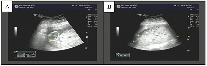
Figure 1. B-ultrasonography at 20 weeks of gestation showing intrauterine singleton and hydatidiform mole tissue. A. Intrauterine single live fetus; B. Large dense dot shaped strong echo area in uterine cavity (167 × 43 × 159mm), indicating partial hydatidiform mole.