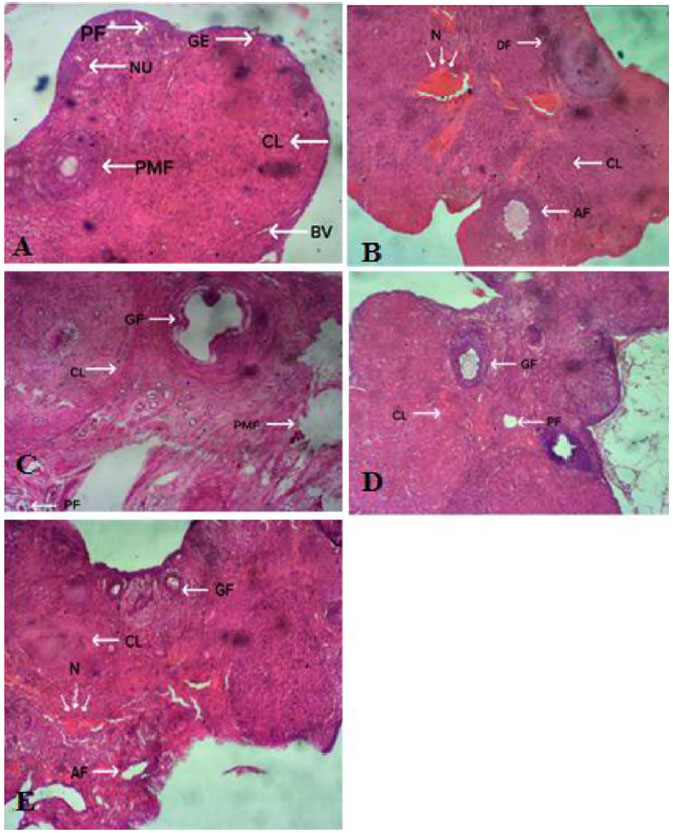
Figure 2. A-E. Photomicrographs of the ovaries of Wistar rats. A. Section of ovaries of rats in Group 1 (Control) showing normal ovarian histology. PF: Primary Follicle; PMF: Primordial Follicle; BV: Blood Vessel; NU: Nucleus; GE: Granulosa Epithelium; CL: Corpus Luteum. B. Section of ovaries of rats in Group 2 (aluminum chloride only) showing N: area of necrosis; CL: Atretic Corpus Luteum; AF: Follicle; DF: Degenerating Follicle. C. Section of ovaries of rats in Group 3 (aluminum chloride + 100mg/kg Xylopia aethiopica) PMF: Primordial follicle; GF: Germinating Follicle. PF: Primary Follicle; CL: Corpus Luteum. Features show a regeneration of follicles and other cells and increased number of Primary Follicles (PF). D. Section of ovaries of rats in Group 4 (aluminum chloride + 50mg/kg Xylopia aethiopica) showing PF: Primary follicle; GF: Germinating Follicle; CL: Corpus Luteum. Features show mild areas of degeneration in the cortex within the follicles. E. Section of ovaries of rats in Group 5 (aluminum chloride + 50mg/kg zinc chloride). AF: Atretic Follicles; GF: Germinating Follicle; CL: Corpus Luteum; N: Necrosis. Features show Antral Follicle (AF) and area of Necrosis. Growing Follicles (GF) are visible.