JBRA Assist. Reprod. 2024;28(2):289-294
ORIGINAL ARTICLE
doi: 10.5935/1518-0557.20240019
Evaluation of the expression level of microRNA-21, microRNA-15a, microRNA-372 in human follicular fluid stem cells-derived oocyte-like cells (OLCs)
1Cellular and Molecular Research Center, Ahvaz Jundishapur University of Medical Sciences, Ahvaz, Iran
2Department of Anatomical Sciences, Faculty of Medicine, Ahvaz Jundishapur University of Medical Sciences, Ahvaz, Iran
3Department of Anatomical Sciences, Faculty of Medicine, Baqiyatallah University of Medical Sciences, Tehran, Iran
4Fertility, Infertility and Perinatology Center, Imam Khomeini Hospital, Ahvaz Jundishapur University of Medical Sciences, Ahvaz, Iran
CONFLICT OF INTEREST
None of the contributing authors has any conflict of interest.
ABSTRACT
Objective: Today, researchers have succeeded in achieving oocyte-like cells through the in vitro differentiation of stem cells. MicroRNAs are key regulators of oocyte development. In this study we decided to evaluate the expression pattern of microRNA-21, microRNA-15a, and microRNA-372 in oocyte-like cells, to determine the maturation stage of oocyte-like cells.
Methods: Human follicular fluid samples were collected and centrifuged, and their cells were divided into 3 groups; day 7 as control group, days 14 and 21. During this period, the cells were evaluated for their morphological appearance and viability by inverted microscopy. RNA isolation was performed and cDNA was reversely transcribed by specific stem-loop RT primers. Real-time RT-PCR was used to detect microRNA expression.
Results: The relative expression of microRNA-21 and microRNA-15a on day 21 was significantly down-regulated compared to the control group (day 7), but microRNA-372 did not show a significant difference. Also, on day 14 compared to the control group (day 7), microRNA-21 did not show a significant difference; but microRNA-15a and microRNA-372 were significantly down-regulated. MicroRNA-21 and microRNA-15a on day 21 compared to day 14 revealed down-regulated levels, but microRNA-372 revealed up-regulated levels.
Conclusions: Our results showed significant decreases in the expression of microRNA-21 and microRNA-15a in oocyte-like cells, as well as in oocytes, which may lead to cytoplasmic maturation, germinal vesicle break down and the completion of meiosis І. In addition, down-regulation expression of microRNA-372 maybe a confirmation that mesenchymal stem cells have differentiated into germ cells, and these cells were differentiated into oocyte-like cells.
Keywords: oocyte-like cells, microRNA-21, microRNA-15a, microRNA-372, infertility, in vitro fertilization
INTRODUCTION
Infertility is one of the main problems couples may face, and it is increasing despite various treatments, at an overall average of 10% in the world (Direkvand-Moghadam et al., 2016). The methods used to treat infertility are called Assisted Reproductive Technology, or ART (Direkvand-Moghadam et al., 2014). In this method, the ovarian follicle is removed from the ovary, and in the laboratory, after separating the oocyte, the follicular fluid is discarded (Astbury et al., 2020). Follicular fluid is an essential component of the ovarian follicle and contains hormones, enzymes, electrolytes and antioxidants for oocyte maturation. Also, the follicular fluid has several types of cells (Taheri Moghadam et al., 2021; Rungsiwiwut et al., 2021).
It has been shown in studies that ovarian follicular fluid-derived cells are stem cells; they also express stem cell markers CD45, CD34, CD90, CD133, SSEA4, OCT4, and NANOG (Rungsiwiwut et al., 2021; Koliakos et al., 2019). Furthermore, researchers demonstrated that follicular fluid stem cells spontaneously differentiate into oocyte-like cells (OLCs) in vitro and express oocyte-specific markers such as Zp2 and Zp3 (Taheri Moghadam et al., 2021; Virant-Klun et al., 2008). According to these data, these cells can be used to investigate the processes of oocyte growth and differentiation during oogenesis in vitro. Oogenesis is controlled by several molecular processes. One of the effective molecules in these processes is microRNAs; which are small, single-stranded, noncoding RNA molecules with 18-24 nucleotides. MicroRNAs bind target mRNA by the RNA-induced silencing complex (RISC) to prevent mRNA translation and protein production; therefore, they play an important role in the posttranscriptional regulation of gene expression in human reproduction (Li et al., 2015; Wahid et al., 2010). The expression of microRNAs in oocyte has been reported in various mammalian species, including human (Xu et al., 2011), bovine (Abd El Naby et al., 2013), mice (Yao et al., 2014), and pig (Hale et al., 2020), and they can control the expression of genes related to oogenesis, including; oocyte development, cytoplasmic maturation (Jenabi et al., 2023), meiotic proteins formation (Liu et al., 2010), and cause the progression of the oocyte from the germinal vesicle stage to meiosis ІІ (Wright et al., 2016).
Up to now, studies investigated the expression pattern of microRNAs in oocyte in vitro maturation (IVM), but there is no report of microRNAs expression in OLCs in vitro. For this reason, to determine the maturation stage of OLCs, we decided to evaluate the expression levels of microRNA-21, microRNA-15a and microRNA-372 in these cells in vitro, so that we can use OLCs to treat female infertility in the future.
METHODS AND MATERIALS
Collection of normal follicular fluid samples
This study was approved by the IRCCS Bioethics Committee (Protocol number IR.AJUMS.MEDICINE.REC.1400.0497). Patients gave their written informed consent on oocyte retrieval day and were informed about FF sample collection. The inclusion criteria included women with no disease and who were referred to the infertility center of Imam Khomeini Hospital in Ahvaz for IVF due to male infertility of their husbands. Exclusion criteria included women with polycystic ovary syndrome (PCOs), premature ovarian failure (POF), thyroid gland disorders, blood pressure disorders, endometriosis and autoimmune diseases. The number of samples included in the study was 12.
Isolation and culture of follicular cells
First, 40-50 ml of human ovarian follicular fluids was collected from each woman who was undergoing IVF therapy during oocyte retrieval through an ultrasound-guided aspiration needle (Lai et al., 2015; Riva et al., 2014). After identifying cumulus-oocyte complexes (COCs) by the first operator for IVF purposes, when no more oocytes were observed by the second operator, follicular fluid (FF) was pooled into a conical 50 ml falcon tube containing two drops of heparin (10-30 IU/ml). We used hypo-osmotic lysis to enrich the follicular cells and the elimination of red blood cells from FF (Lobb & Younglai, 2006). In this technique, follicular aspirates were centrifuged at 800g for 30 min. Then, aspirating supernatant was collected and the cell slurry was transferred into a 15 ml falcon tube. In the next step, 9.0 ml of the sterile distilled water was poured into the cell slurry, and the tube was blended. After 20s, 1.0 ml of 10× concentrated phosphate-buffered saline (PBS, pH = 7.2) was added and mixed in the tube. The tubes were centrifuged at 400g for 10 min again. Finally, the cell pellet was resuspended in 0.5 ml of Dulbecco’s modified Eagle’s medium (DMEM; Sigma-Aldrich). Cell counting and viability test were performed on aliquots in 0.2% trypan blue by a hemocytometer (Lobb & Younglai, 2006). FF-aspirated cells were seeded onto four-well plates (BD Biosciences) at 1 × 10⁵ cells concentration. The growing cells were separated in a DMEM supplemented with 10% fetal bovine serum (FBS; Gibco-BRL) and 1% penicillin/streptomycin (Gibco). Non-adherent cells were thrown away after 48h; the cultures were kept at 37°C in a 5% CO2 humidified atmosphere and monitored daily. The culture medium was replaced every 2 days. The samples were divided into three groups: day 7, day 14 and day 21. The cells were monitored daily through an inverted microscope (Nikon, Japan) for morphology assessment. Every two days, the cell culture medium was changed. The size of cells was measured by software: Analysis LS Research 3.2 (Olympus company) (Taheri Moghadam et al., 2021).
RNA extraction and cDNA synthesis
The extraction of total RNA was conducted from 4-6106 using the SinaPure RNA Mini Kit (SinaClon). According to the manufacturer′s instruction. First, lysis buffer and precipitation solution were added into each sample. After that, all the samples were centrifuged and they were washed twice with wash buffer, centrifugation was done again. Finally, RNA samples were eluted in a 50 ml of RNase-free water. The quality of the extracted RNA sample was determined by the NanoDrop™ 2000c spectrophotometer at an optical density of 260 to 280, which is the best case, close to 2, and it was 1.98 in the extracted samples. Later, 500ng of total RNAs underwent reverse-transcription into complementary DNA using the cDNA synthesis kit (SinaClon), which include dNTP, gene-specific primer, M-MuLV Reverse Transcriptase, 5x Buffer M-MuLV and RNase inhibitor. Then, all the samples were left for 50 minutes at 50°C, ending in 15 minute-cycles at 70°C in an eppendorf Master Cycler. Finally, all the samples were stored at -20°C prior to RNA purification (Zhang et al., 2007).
Quantitative RT-PCR
The real-time method was used to quantitatively check the expression of microRNAs. The volume of the real-time mix for each reaction included: 10µl Real Qplus 2x Mastermix Green (Ampliqon, Denmark), 2 µl cDNA, 1µl primer reverse, 1µl primer forward, 6µl distilled water and each sample was analyzed in duplicate. The temperature program of the real-time device (ABI 1500 PCR) in each cycle included: initial denaturation 95°C for 15 minutes, denaturation 95◦C for 20 seconds, annealing 60°C for 20 seconds, extension 72°C for 20 seconds. There were 40 cycles and the expression of U6 was used as a housekeeping control gene (Naji et al., 2018). The sequence of the primers used in this research is according to Table 1.
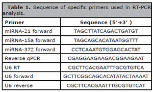
Table 1. Sequence of specific primers used in RT-PCR analysis.
Statistical analysis
The formula 2-∆∆Ct was used to calculate the relative expression of genes. Statistical analysis was done by using the independent-sample T-test and one-way ANOVA. The Graph Pad Prism6 (Graph Pad Software Inc., San Diego CA, USA) was used to draw the graphs. P value ≤ 0.05 was considered statistically significant (Xu et al., 2011).
RESULTS
Morphology of normal follicular fluid cells in vitro
The isolated cells on the first day of cultivation had a round morphology (Figure 1a). After growing the adherent small mesenchymal cells, they attached to the bottom of the 4-well plate and formed a cell colony over time, and fibroblast-like cells appeared in between these cell colonies (Figure 1b). On day 7 of the culture, oocyte-like cells appeared in the medium; they had a size of 20µm and appeared next to epithelial cells and fibroblast-like cells in the environment (Figure 1c). Gradually, over time, OLCs increased in size, so that, on day 14 of the culture, they reached a size of about 25 µm (Figure 1d), on day 17 of culture, the size of the cells was 27 µm, and finally on day 21, they became larger and reached the size of 33 µm, and they were located next to fibroblast-like cells and the cell colonies (Figure 1e, f).
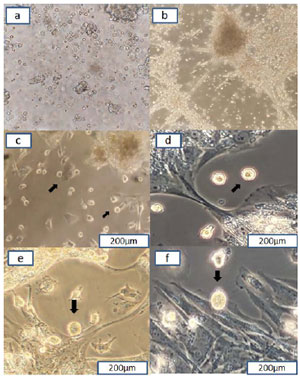
Figure 1. Cells derived from follicular fluid on the first day of culture (a). On day 3 of culture and cell colony formation and appearance of fibroblast-like cells (b). On day7, OLCs next to epithelial and fibroblast-like cells(c). On day 14 of culture (d). On day 17 of culture (e). On day 21 of culture, OLCs (f). Magnification 20x; arrows point to the OLCs.
Real-time PCR results of microRNA gene expression analysis
Expression of microRNA-21
As shown in Figure 2, the expression of microRNA-21 on day 21 was significantly down-regulated compared to that of the control group (day 7) (p=0.0005, 0.1-fold), but on day 14 compared to the control group, there was no significant difference (p=0.24). Also, expression of microRNA-21 on day 21 compared to day 14 was significantly down-regulated (p=0.0022, ~ 0.1-fold).
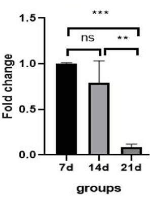
Figure 2. Expression of miRNA-21 gene in OLCs in control group (day 7) and study groups (day 14 and day 21). ***: p=0.0005, **: p=0.0022, ns = not significant.
Expression of microRNA-15a
As shown in Figure 3, the expression of microRNA-15a on day 14 was significantly down-regulated compared to the control group (p=0.0001, 0.25-fold). Also on day 21, compared to the control group, it was significantly down-regulated (p=0.0001, 0.1-fold). On day 21 compared to day 14, the expression of microRNA-15a was significantly down-regulated (p=0.0019, 0.4-fold).
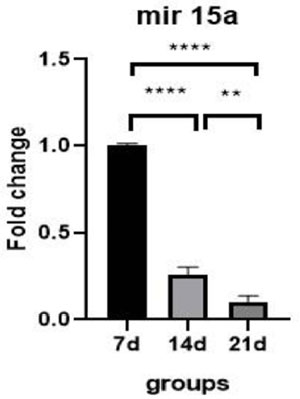
Figure 3. Expression miRNA-15a gene in OLCs in control groups (day 7) and study groups (day 14 and day 21). **: p=0.0019, ****: p=0.0001.
Expression of microRNA-372
As shown in Figure 4, the expression of microRNA-372 on day 14 was significantly down-regulated compared to the control group (p=0.0002, 0.46-fold), but on day 21 compared to the control group did not show a significant difference (p=0.42). On day 21 compared to day 14, the expression of microRNA-372 was significantly up-regulated (p=0.0001, 2-fold).
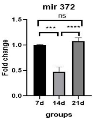
Figure 4. Expression of miRNA-372 gene in OLCs in control groups (day 7) and study groups (day 14 and day 21). ***: p=0.0002, ****: p=0.0001, ns=not significant.
DISCUSSION
The process of oocyte maturation includes dynamic epigenic mechanisms and bidirectional interaction between the oocyte and surrounding somatic cells. As a result, it leads to progress in the molecular processes related to folliculogenesis and eventually, development of the oocyte from meiosis І to meiosis ІІ. One of the key regulators of these processes are microRNAs, which play an important role in fertility. According to the studies, microRNAs had different expression patterns in different stages of oocyte development, including meiosis І, meiosis ІІ and germinal vesicle break down (GVBD) stage. In this study, for the first time, we investigated the expression level of microRNAs in oocyte-like cells in order to find out in which differentiation stage these cells were. Maybe these cells can be used as substitutes for oocytes in the treatment of women whose infertility problem is due to lack of oocytes or ineffective oocytes (Maalouf et al., 2016; Hossain et al., 2012; Gilchrist et al., 2016)
In this study, cells isolated from follicular fluid on the first day had a round shape, then, cell colonies were gradually formed, and a heterogeneous population of epithelial-like fibroblast-granulosa cells appeared in the medium on day 7. According to previous studies, these cells have the ability to express mesenchymal stem cell markers, including OCT4, NANOG, and SSEA4, so they can differentiate into different cell lines, including cartilage cells, adipose cells, and bone cells. Also, they are able to differentiate into germ cells and express DDX4, which is a germ cell marker. Therefore, these cells are mesenchymal stem cells (Rungsiwiwut et al., 2021; Koliakos et al., 2019).
One of the characteristics of oocyte maturation is the presence of zona pellucida (ZP) glycoprotein around the cell, which is a type of extracellular matrix and consists of 4 types. This protein has an important role in the development of the oocyte during the process of oogenesis, the interaction between sperm and egg, and the development of the embryo during fertility in women (Wassarman & Litscher, 2022). In the present study, on day 7, the stem cells were differentiated into round and small cells, which gradually increased in size over time. Previous studies have indicated that these cells are able to express oocyte surface proteins, which are ZP2 and ZP3 (Taheri Moghadam et al., 2021; Azandeh et al., 2022). As a result, they are oocyte-like cells.
In the present study, OLCs were 20μm in size on day 7. Gradually, their size increased so that they were 25μm on day 14 and 33μm on day 21. These results are also consistent with Lai’s study. Lai et al. (2015) showed that human follicular fluid epithelial cells formed cell colonies in the culture medium, and after some time they differentiated into oocyte-like cells, and these cells during the culture period from the second week to the fourth week had different sizes.
In our study, for the first time, we investigated the expression of microRNA-15a, microRNA-21, and microRNA-372 in oocyte-like cells derived from normal human follicular fluid stem cells. In this context, studies show that microRNAs play an important role in regulating cellular processes, including cell differentiation, apoptosis, and cell division (Trohatou et al., 2014). One of the microRNAs investigated in this study was microRNA-21. MicroRNA-21 has a regulatory role in many biological activities of the cell; therefore, it is considered a biomarker for the diagnosis of diseases including cancer and heart diseases (Jenabi et al., 2023; Jenike & Halushka, 2021). MicroRNA-21 also plays a vital role in the differentiation of mesenchymal stem cells, and its inhibition causes a decrease in the differentiation of stem cells (Sekar et al., 2015). Dehghan et al. (2021) conducted a study with the aim of evaluating the effect of microRNA-21 on the development process of mouse oocytes and their blastocysts in in vitro maturation (IVM). They showed that the increase in the expression of microRNA-21 in cumulus cells decreased the progress of mitosis, decreased the amount of Glutathione-S-transferase (GSH), and the expression of BMPR2 and PTX3 genes in oocytes. On the other hand, in oocytes, decreasing the expression of microRNA-21 increased cytoplasmic maturation, but it had no effect on the maturation of the oocyte nucleus or its progress from the germinal vesicle stage to meiosis ІІ.
Also, in this study, we investigated the relationship between the expression level of microRNA-21 and the OCT4 factor. They observed that the expression level of the OCT4 factor was significantly reduced in the blastocysts, which induced the microRNA-21 silencing factor (Dehghan et al., 2021). As we know, OCT4 is a pluripotency factor in mesenchymal stem cells. In the study of Azandeh et al. (2022), the expression level of this factor during the culture of normal human follicular fluid stem cells has gradually decreased, and this indicated the differentiation of stem cells into oocyte-like cells and the reduction of stem cells in the culture medium. In our study there was a significant down-regulation in microRNA-21 expression during the culture in the second and third weeks. These changes are consistent with the study of Dehghan et al. (2021), and similar to the expression pattern of microRNA-21 in mouse oocytes. It is also possible that the decrease in the expression of microRNA-21 and OCT4 factor in the culture medium indicates the differentiation of stem cells into oocyte-like cells, which according to Dehghan’s study (Dehghan et al., 2021), maybe oocyte-like cells are like oocyte in the cytoplasmic maturation stage.
Another microRNA studied was microRNA-15a, which belongs to the microRNA-15/16 cluster. This class is known as a tumor suppressor and works by targeting the BCL2, MCL1, CCND1, and WNT3A genes. As a result, by affecting the cell cycle of cancer cells, they reduce cell proliferation, promote apoptosis and suppress tumors (Aqeilan et al., 2010). In 2011, Xu et al. investigated microRNAs expression during human oocytes meiosis in vitro. They showed that the expression level of microRNA-15a was significantly down-regulated during the development process of human oocytes from the germinal vesicle stage to meiosis ІІ (Xu et al., 2011). In our study, on days 14 and 21, compared to the day 7, the expression level of microRNA-15a was significantly down-regulated. These changes are consistent with the study of Xu et al., and similar to the expression pattern of microRNA-15a in human oocytes. As a result, according to the study by Xu et al. (2011), maybe oocyte-like cells are in GVBD and completion of meiosis I.
MicroRNA-372 was another microRNA investigated in this study, which has an anti-oncogenic role in tumors involving germ cells, ovarian cancer, gastric adenocarcinoma, and liver cells. MicroRNA-372 plays its role through proteins participating in apoptosis and the cell cycle, thus reducing cell proliferation and promoting apoptosis (Guan et al., 2017). Tran et al. (2016) showed that microRNA-372 increases during the differentiation of stem cells into primitive germ cells. This researcher reported that microRNA-372 can increase primordial germ cells by suppressing multiple cell cycle pathways as well as epigenetic processes (Tran et al., 2016). On the other hand, Rungsiwiwut et al. (2021) showed that follicular fluid stem cells can differentiate into germ cells and express the DDX4 marker. In our study, the expression level of microRNA-372 was significantly up-regulated on day 21 compared to day 14, which may indicate that in the second week (day 14), follicular fluid stem cells are more differentiated into germ cells. But the expression level of microRNA-372 was significantly down-regulated on day 14 compared to day 7; maybe it was because the stem cells were more differentiated into OLCs during the first and second weeks.
The results of the present study showed that, the expression pattern of microRNA-21 and microRNA-15a in oocyte-like cells is similar to the expression pattern of these microRNAs in the oocyte. As a result, it can be said that the OLCs in the present study have probably progressed to the stage of GVBD and the completion of meiosis І; and cytoplasmic maturation has taken place simultaneously with these changes. Also, the significant reduction of microRNA-372 was a confirmation that mesenchymal stem cells (MSCs) have differentiated into germ cells. Then these germ cells were differentiated into OLCs, but confirmation of this issue requires further investigations.
ACKNOWLEDGMENT
This study was supported by the Cellular and molecular Research Center, Ahvaz Jundishapur University of Medical Sciences, Ahvaz, Iran (grant number IR.AJUMS.MEDICINE.REC.1400.0497).We appreciate the assistance of the laboratory technicians at the IVF clinic of Imam Hospital (Mrs. Pour shamsa and Lami, F.) for helping us in collecting the samples. This study is the M.Sc. thesis of Ms. Shahrooie
REFERENCES
Abd El Naby WS, Hagos TH, Hossain MM, Salilew-Wondim D, Gad AY, Rings F, Cinar MU, Tholen E, Looft C, Schellander K, Hoelker M, Tesfaye D. Expression analysis of regulatory microRNAs in bovine cumulus oocyte complex and preimplantation embryos. Zygote. 2013;21:31-51. PMID: 22008281 DOI: 10.1017/S0967199411000566 Medline
Aqeilan RI, Calin GA, Croce CM. miR-15a and miR-16-1 in cancer: discovery, function and future perspectives. Cell Death Differ. 2010;17:215-20. PMID: 19498445 DOI: 10.1038/cdd.2009.69 Medline
Astbury P, Subramanian GN, Greaney J, Roling C, Irving J, Homer HA. The Presence of Immature GV- Stage Oocytes during IVF/ICSI Is a Marker of Poor Oocyte Quality: A Pilot Study. Med Sci. 2020;8:4. PMID: 31963146 DOI: 10.3390/medsci8010004 Medline
Azandeh S, Moghadam MT, Rashno M, Zargar M, Zadeh PA. The Effect of Different Doses of Melatonin on in Vitro Maturation of Human Follicular Fluid-Derived Oocyte-Like Cells. JBRA Assist Reprod. 2022;26:288-98 PMID: 34786903 DOI: 10.5935/1518-0557.20210086 Medline
Dehghan Z, Mohammadi-Yeganeh S, Rezaee D, Salehi M. MicroRNA-21 is involved in oocyte maturation, blastocyst formation, and pre-implantation embryo development. Dev Biol. 2021;480:69-77. PMID: 34411594 DOI: 10.1016/j.ydbio.2021.08.008 Medline
Direkvand-Moghadam A, Delpisheh A, Direkvand-Moghadam A. Effect of infertility on the quality of life, a cross-sectional study. J Clin Diagn Res. 2014;8:OC13-5.PMID: 25478412 DOI: 10.7860/JCDR/2014/8481.5063. Medline
Gilchrist GC, Tscherner A, Nalpathamkalam T, Merico D, LaMarre J. MicroRNA Expression during Bovine Oocyte Maturation and Fertilization. Int J Mol Sci. 2016;17:396. PMID: 26999121 DOI: 10.3390/ijms1703039 Medline
Guan X, Zong ZH, Chen S, Sang XB, Wu DD, Wang LL, Liu Y, Zhao Y. The role of miR-372 in ovarian carcinoma cell proliferation. Gene. 2017;624:14-20. PMID: 28456593 DOI: 10.1016/j.gene.2017.04.043 Medline
Hale BJ, Li Y, Adur MK, Ross JW. Inhibition of germinal vesicle breakdown using IBMX increases microRNA-21 in the porcine oocyte. Reprod Biol Endocrinol. 2020;18:39. PMID: 32393269 DOI: 10.1186/s12958-020-00603-1 Medline
Hossain MM, Salilew-Wondim D, Schellander K, Tesfaye D. The role of microRNAs in mammalian oocytes and embryos. Anim Reprod Sci. 2012;134:36-44. PMID: 22921265 DOI: 10.1016/j.anireprosci.2012.08.009 Medline
Jenabi M, Khodarahmi P, Tafvizi F, Bostanabad SZ. Evaluation of the potential of miR-21 as a diagnostic marker for oocyte maturity and embryo quality in women undergoing ICSI. Sci Rep. 2023;13:1440. PMID: 36697494 DOI: 10.1038/s41598-023-28686-x Medline
Jenike AE, Halushka MK. miR-21: a non-specific biomarker of all maladies. Biomark Res. 2021;9:18. PMID: 33712063 DOI: 10.1186/s40364-021-00272-1 Medline
Lai D, Xu M, Zhang Q, Chen Y, Li T, Wang Q, Gao Y, Wei C. Identification and characterization of epithelial cells derived from human ovarian follicular fluid. Stem Cell Res Ther. 2015;6:13. PMID: 25889077 DOI: 10.1186/s13287-015-0004-6 Medline
Li Y, Fang Y, Liu Y, Yang X. MicroRNAs in ovarian function and disorders. J Ovarian Res. 2015;8:51. PMID: 26232057 DOI: 10.1186/s13048-015-0162-2 Medline
Liu HC, Tang Y, He Z, Rosenwaks Z. Dicer is a key player in oocyte maturation. J Assist Reprod Genet. 2010;27:571-80. PMID: 20827505 DOI: 10.1007/s10815-010-9456-x Medline
Lobb DK, Younglai EV. A simplified method for preparing IVF granulosa cells for culture. J Assist Reprod Genet. 2006;23:93-5. PMID: 16552632 DOI: 10.1007/s10815-006-9025-5 Medline
Maalouf SW, Liu WS, Pate JL. MicroRNA in ovarian function. Cell Tissue Res. 2016;363:7-18. PMID: 26558383 DOI: 10.1007/s00441-015-2307-4 Medline
Naji M, Nekoonam S, Aleyasin A, Arefian E, Mahdian R, Azizi E, Shabani Nashtaei M, Amidi F. Expression of miR-15a, miR-145, and miR-182 in granulosa-lutein cells, follicular fluid, and serum of women with polycystic ovary syndrome (PCOS). Arch Gynecol Obstet. 2018;297:221-31. PMID: 29071578 DOI: 10.1007/s00404-017-4570-y Medline
Riva F, Omes C, Bassani R, Nappi RE, Mazzini G, Icaro Cornaglia A, Casasco A. In-vitro culture system for mesenchymal progenitor cells derived from waste human ovarian follicular fluid. Reprod Biomed Online. 2014;29:457-69. PMID: 25131558 DOI: 10.1016/j.rbmo.2014.06.006 Medline
Rungsiwiwut R, Numchaisrika P, Thuwanut P, Pruksananonda K. Characterization of stem cells from human ovarian follicular fluid; a potential source of autologous stem cell for cell-based therapy. Hum Cell. 2021;34:300-9. PMID: 33543452 DOI: 10.1007/s13577-020-00439-2 Medline
Sekar D, Saravanan S, Karikalan K, Thirugnanasambantham K, Lalitha P, Islam VI. Role of microRNA 21 in mesenchymal stem cell (MSC) differentiation: a powerful biomarker in MSCs derived cells. Curr Pharm Biotechnol. 2015;16:43-8. PMID: 25564252 DOI: 10.2174/138920101601150105100851 Medline
Taheri Moghadam M, Saki G, Nikbakht R, Eftekhari AR Moghadam. Bone morphogenetic protein 15 induces differentiation of mesenchymal stem cells derived from human follicular fluid to oocyte-like cell. Cell Biol Int. 2021;45:127-39. PMID: 32997425 DOI: 10.1002/cbin.11475 Medline
Tran ND, Kissner M, Subramanyam D, Parchem RJ, Laird DJ, Blelloch RH. A miR-372/let-7 Axis Regulates Human Germ Versus Somatic Cell Fates. Stem Cells. 2016;34:1985-91. PMID: 27066911 DOI: 10.1002/stem.2378 Medline
Trohatou O, Zagoura D, Bitsika V, Pappa KI, Antsaklis A, Anagnou NP, Roubelakis MG. Sox2 suppression by miR-21 governs human mesenchymal stem cell properties. Stem Cells Transl Med. 2014;3:54-68. PMID: 24307698 DOI: 10.5966/sctm.2013-0081 Medline
Virant-Klun I, Zech N, Rozman P, Vogler A, Cvjeticanin B, Klemenc P, Malicev E, Meden-Vrtovec H. Putative stem cells with an embryonic character isolated from the ovarian surface epithelium of women with no naturally present follicles and oocytes. Differentiation. 2008;76:843-56. PMID: 18452550 DOI: 10.1111/j.1432-0436.2008.00268.x Medline
Wahid F, Shehzad A, Khan T, Kim YY. MicroRNAs: synthesis, mechanism, function, and recent clinical trials. Biochim Biophys Acta. 2010;1803:1231-43. PMID: 20619301 DOI: 10.1016/j.bbamcr.2010.06.013 Medline
Wassarman PM, Litscher ES. Female fertility and the zona pellucida. Elife. 2022;11:e76106. PMID: 35076396 DOI: 10.7554/eLife.76106 Medline
Wright EC, Hale BJ, Yang CX, Njoka JG, Ross JW. MicroRNA-21 and PDCD4 expression during in vitro oocyte maturation in pigs. Reprod Biol Endocrinol. 2016;14:21. PMID: 27084064 DOI: 10.1186/s12958-016-0152-2 Medline
Xu YW, Wang B, Ding CH, Li T, Gu F, Zhou C. Differentially expressed micoRNAs in human oocytes. J Assist Reprod Genet. 2011;28:559-66. PMID: 21647640 DOI: 10.1007/s10815-011-9590-0 Medline
Yao G, Liang M, Liang N, Yin M, Lü M, Lian J, Wang Y, Sun F. MicroRNA-224 is involved in the regulation of mouse cumulus expansion by targeting Ptx3. Mol Cell Endocrinol. 2014;382:244-53. PMID: 24145127 DOI: 10.1016/j.mce.2013.10.014 Medline
Zhang HH, Wang XJ, Li GX, Yang E, Yang NM. Detection of let-7a microRNA by real-time PCR in gastric carcinoma. World J Gastroenterol. 2007;13:2883-8. PMID: 17569129 DOI: 10.3748/wjg.v13.i20.2883 Medline