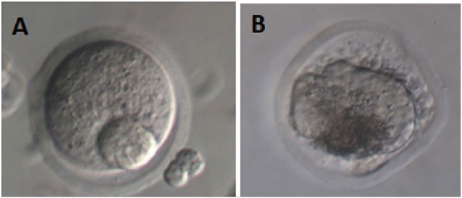
Figure 4. Illustration of oocytes recovered from follicles at D11. A: intact oocyte, B: atretic oocyte. A photomicrograph was obtained using an inverted microscope (Olympus MII, Japan, 400x, Hoffman Modulation Contrast). Source: Own authorship.
Figure 4. Illustration of oocytes recovered from follicles at D11. A: intact oocyte, B: atretic oocyte. A photomicrograph was obtained using an inverted microscope (Olympus MII, Japan, 400x, Hoffman Modulation Contrast). Source: Own authorship.