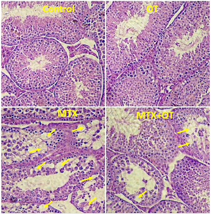
Figure 2. Light microscopy of testicular tissue from the control and experimental groups (Arrows show vacuoles in the germinal epithelium); H&E staining; Magnifications: ×250.
Figure 2. Light microscopy of testicular tissue from the control and experimental groups (Arrows show vacuoles in the germinal epithelium); H&E staining; Magnifications: ×250.